Home
Understanding the Electrocardiogram

Main navigation

  • Home
  • Understanding ECGs
    • Introduction
User account menu
  • Log in

Breadcrumb

  1. Home
  2. Understanding the Electrocardiogram In Health & Disease
  3. 8.0.0 The Bradycardias frame i-introduction
  4. 8.2.0 SA Block (frame 41)

8.2.15 (56)

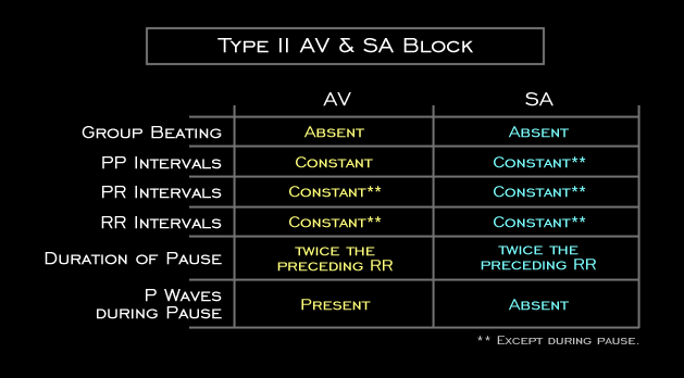
In this table, the electrocardiographic features of type II 2nd degree AV and SA block are compared. Note that the only difference between the two is the presence of P waves during the pause with AV block and their absence with SA block.

Book traversal links for 8.2.15 (56)

  • 8.2.14 (55)
  • Up
  • 8.3.0 Group Beating (frame 57)

Site is under construction

Book navigation

  • Introduction to First Edition
  • 1.0.0 Generation of the ECG
  • 2.0.0 The Normal Electrocardiogram
  • 3.0.0 Inter and intra-ventricular Conduction Disturbances
  • 4.0.0 Ventricular Hypertrophy
  • 5.0.0 ELECTROLYTE ABNORMALITIES, DRUG EFFECTS AND THE LONG QT SYNDROMES
  • 6.0.0 Ischemia and Infarction - Introduction (frame i and ii)
  • 7.0.0 Tachycardias- Introduction
  • 8.0.0 The Bradycardias frame i-introduction
    • 8.1.0 Sinus Bradycardia and AV block (frame 1)
    • 8.2.0 SA Block (frame 41)
      • 8.2.1 (42)
      • 8.2.2 (43)
      • 8.2.3(44)
      • 8.2.4 (45)
      • 8.2.5 (46)
      • 8.2.6 (47)
      • 8.2.7 (48)
      • 8.2.8 (49)
      • 8.2.9 (50)
      • 8.2.10 (frame 51)
      • 8.2.11 (52)
      • 8.2.12 (53)
      • 8.2.13 (54)
      • 8.2.14 (55)
      • 8.2.15 (56)
    • 8.3.0 Group Beating (frame 57)
  • 9.0.0 The ECG of Heart Murmurs-introduction
  • 10.0.0 The Electrocardiogram in the Emergency Department-Introduction
Powered by Drupal