
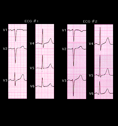
A change in the position of the chest leads, particularly leads V2, V3 and V4 will change the relationship of these leads to the heart and result in a change in the QRS complex as recorded by these leads. The ECGs shown here were recorded on two successive days from a 67 year old patient with no known or suspected heart disease. On the first day (ECG #1), there is normal R wave progression in leads V1-V3 and the tracing is normal. On the next day (ECG #2), there is poor R wave progression with a very small R wave in leads V1-V3 and more subtle changes in leads V4-6. These changes indicate that on day 2, the relationship of the heart to the chest wall electrodes had changed. A change in the patient’s position at the time of the recording (such as sitting or turned to one side) or a change in the position of the chest electrodes, particularly those placed in the V3 and V4 positions would account for the change in the ECG. If ECG #2 were the only tracing available, the poor R wave progression in leads V1-V3 might have been interpreted as suggesting a prior anterior wall infarction.