Home
Understanding the Electrocardiogram

Main navigation

  • Home
  • Understanding ECGs
    • Introduction
User account menu
  • Log in

Breadcrumb

  1. Home
  2. Understanding the Electrocardiogram In Health & Disease
  3. 6.0.0 Ischemia and Infarction - Introduction (frame i and ii)
  4. 6.2.0 Q WAVES (frame 53)

6.2.8 (61)

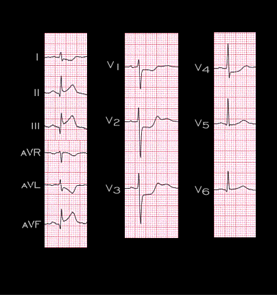
As discussed earlier (see page 6.1.37), posterior wall involvement is difficult to recognize in the standard 12 lead electrocardiogram because recording electrodes are not routinely placed on the posterior chest wall. Ischemia and infraction of the posterior wall do cause changes in leads V1 and V2, the most anteriorly placed chest leads, that are the opposite, or reciprocal of the changes occurring posteriorly and likely to be recorded by posterior chest wall leads, V8 and V9. The ST segment depression in leads V1 and V2 in this ECG from the patient with the inferior and posterior wall infarction that was shown earlier on page 6.1.38, reflects the posterior wall involvement. Note also the R waves in leads V1 and V2. These are slightly wider than those that normally occur in these leads and may be the reciprocal of Q waves that would have been recorded in leads placed in the V8 and V9 positions.

Book traversal links for 6.2.8 (61)

  • 6.2.7
  • Up
  • 6.2.9 (64)

Site is under construction

Book navigation

  • Introduction to First Edition
  • 1.0.0 Generation of the ECG
  • 2.0.0 The Normal Electrocardiogram
  • 3.0.0 Inter and intra-ventricular Conduction Disturbances
  • 4.0.0 Ventricular Hypertrophy
  • 5.0.0 ELECTROLYTE ABNORMALITIES, DRUG EFFECTS AND THE LONG QT SYNDROMES
  • 6.0.0 Ischemia and Infarction - Introduction (frame i and ii)
    • 6.1.0 ST Segment (frame1)
    • 6.2.0 Q WAVES (frame 53)
      • 6.2.1 (54)
      • 6.2.2 (55)
      • 6.2.3 (56)
      • 6.2.4 (57)
      • 6.2.5 (58)
      • 6.2.6 (59)
      • 6.2.7
      • 6.2.8 (61)
      • 6.2.9 (64)
      • 6.2.10 (68)
      • 6.2.11 (69)
      • 6.2.12 (frame 70)
      • 6.2.13(71)
      • 6.2.14 (72)
      • 6.2.15 (73)
      • 6.2.16 (74)
    • 6.3.0 T WAVES (frame 75)
    • 6.4.0 (Bundle Branch Blocks and othe IVCDs--frame 87)
    • 6.5.0 (frame 106) FALSE POSITIVES
  • 7.0.0 Tachycardias- Introduction
  • 8.0.0 The Bradycardias frame i-introduction
  • 9.0.0 The ECG of Heart Murmurs-introduction
  • 10.0.0 The Electrocardiogram in the Emergency Department-Introduction
Powered by Drupal