
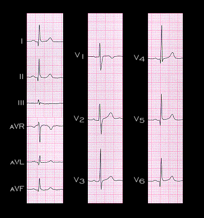
Sometimes, however, tall R waves in leads V1 and V2 may be present and still within normal limits even in the absence of a posterior wall infarction or right ventricular hypertrophy.
The ECG shown here is from a 39 year woman who had no evidence of coronary artery disease and no evidence of right ventricular hypertrophy. The narrow Q waves in leads I, II, aVL, V4, V5 and V6 are the normal “septal” Q waves. The prominent R wave in leads V1 is equal in amplitude to the S wave and is at the upper limit of normal. An interpretation suggesting either possible right ventricular hypertrophy or an infarction involving the posterior and lateral walls would be understandable, but not necessarily correct.