Home
Understanding the Electrocardiogram

Main navigation

  • Home
  • Understanding ECGs
    • Introduction
User account menu
  • Log in

Breadcrumb

  1. Home
  2. Understanding the Electrocardiogram In Health & Disease
  3. 3.0.0 Inter and intra-ventricular Conduction Disturbances
  4. 3.4.0 Aberrant Ventricular Conduction (52)

3.4.12 (65)

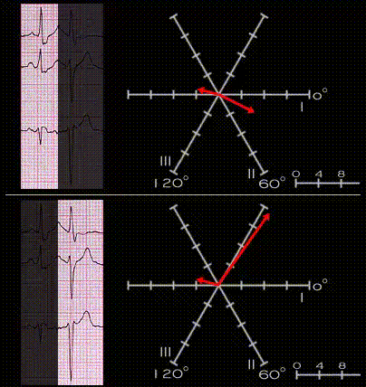
[NEED TO ADD LEADS aVR, AVL and aVF, AND A SMALL INITIAL VECTOR TO THE FIGURE AND THEN ALSO CORRECT THE LOOP IN THE LOWER FRAME] The upper loop represents the non-premature beat. It shows a small initial vector with an axis of +60 degrees, (the small initial R in 3 and aVF), the main vector with an axis of +30 degrees and the terminal vector which represents the right bundle branch block with an axis of +200 degrees. The lower loop represents the premature beat. The initial small vector and the terminal right bundle branch block vector are unchanged, but the main QRS vector has shifted to the left with an axis of -60 degrees.

Book traversal links for 3.4.12 (65)

  • 3.4.11 (64)
  • Up
  • 3.4.13 (68)

Site is under construction

Book navigation

  • Introduction to First Edition
  • 1.0.0 Generation of the ECG
  • 2.0.0 The Normal Electrocardiogram
  • 3.0.0 Inter and intra-ventricular Conduction Disturbances
    • 3.0.1 Introduction Continued
    • 3.1.0 Right Bundle Branch Block (RBBB)
    • 3.2.0 Left Bundle Branch Block (LBBB)
    • 3.3.0 Fascicular blocks (frame 29)
    • 3.4.0 Aberrant Ventricular Conduction (52)
      • 3.4.1 (53)
      • 3.4.2 (54)
      • 3.4.3 (55)
      • 3.4.4 (56)
      • 3.4.5 (57)
      • 3.4.6 (58)
      • 3.4.7 (59)
      • 3.4.8.(THIS IS FRAME 61)
      • 3.4.9 (62)
      • 3.4.10 (63)
      • 3.4.11 (64)
      • 3.4.12 (65)
      • 3.4.13 (68)
      • 3.4.14 (69)
      • 3.4.15 (frame 75)
      • 3.4.16 (76)
    • 3.5.0 Ventricular Pre-excitation (77)
  • 4.0.0 Ventricular Hypertrophy
  • 5.0.0 ELECTROLYTE ABNORMALITIES, DRUG EFFECTS AND THE LONG QT SYNDROMES
  • 6.0.0 Ischemia and Infarction - Introduction (frame i and ii)
  • 7.0.0 Tachycardias- Introduction
  • 8.0.0 The Bradycardias frame i-introduction
  • 9.0.0 The ECG of Heart Murmurs-introduction
  • 10.0.0 The Electrocardiogram in the Emergency Department-Introduction
Powered by Drupal