Home
Understanding the Electrocardiogram

Main navigation

  • Home
  • Understanding ECGs
    • Introduction
User account menu
  • Log in

Breadcrumb

  1. Home
  2. Understanding the Electrocardiogram In Health & Disease
  3. 3.0.0 Inter and intra-ventricular Conduction Disturbances
  4. 3.4.0 Aberrant Ventricular Conduction (52)

3.4.9 (62)

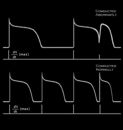
In the figure shown here, the last beat in both the upper and lower panels is premature and their coupling intervals are the same. In the upper panel, the basic rate is slower than in the lower panel. As a result, the duration of the non-premature action potentials is greater than in the lower panel, where the basic rate is faster. Because of this, the premature action potential shown in the upper panel arises from an incompletely repolarized fiber and a membrane potential that is less negative than the resting potential. Its upstroke velocity is reduced because of the voltage dependency of the sodium current and it will conduct aberrantly. The premature action potential in the lower panel, where the basic rate is faster, arises from a completely repolarized fiber. Its upstroke velocity is not reduced and it will conduct normally, without aberration.

Book traversal links for 3.4.9 (62)

  • 3.4.8.(THIS IS FRAME 61)
  • Up
  • 3.4.10 (63)

Site is under construction

Book navigation

  • Introduction to First Edition
  • 1.0.0 Generation of the ECG
  • 2.0.0 The Normal Electrocardiogram
  • 3.0.0 Inter and intra-ventricular Conduction Disturbances
    • 3.0.1 Introduction Continued
    • 3.1.0 Right Bundle Branch Block (RBBB)
    • 3.2.0 Left Bundle Branch Block (LBBB)
    • 3.3.0 Fascicular blocks (frame 29)
    • 3.4.0 Aberrant Ventricular Conduction (52)
      • 3.4.1 (53)
      • 3.4.2 (54)
      • 3.4.3 (55)
      • 3.4.4 (56)
      • 3.4.5 (57)
      • 3.4.6 (58)
      • 3.4.7 (59)
      • 3.4.8.(THIS IS FRAME 61)
      • 3.4.9 (62)
      • 3.4.10 (63)
      • 3.4.11 (64)
      • 3.4.12 (65)
      • 3.4.13 (68)
      • 3.4.14 (69)
      • 3.4.15 (frame 75)
      • 3.4.16 (76)
    • 3.5.0 Ventricular Pre-excitation (77)
  • 4.0.0 Ventricular Hypertrophy
  • 5.0.0 ELECTROLYTE ABNORMALITIES, DRUG EFFECTS AND THE LONG QT SYNDROMES
  • 6.0.0 Ischemia and Infarction - Introduction (frame i and ii)
  • 7.0.0 Tachycardias- Introduction
  • 8.0.0 The Bradycardias frame i-introduction
  • 9.0.0 The ECG of Heart Murmurs-introduction
  • 10.0.0 The Electrocardiogram in the Emergency Department-Introduction
Powered by Drupal