
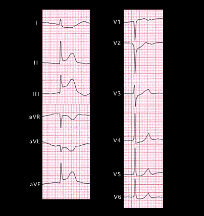
The ECG shown here is from a 72 year old patient with chest pain who was taken to the emergency department by the EMT squad. It shows changes of an acute inferior wall infarction with ST elevation in leads II, III, and aVF. Note that there is also slight ST elevation in lead V1, that the elevation of the ST segment is greater in lead III than in lead II and that there is slight ST depression in leads 1, aVL and leads V2-6. These ST segment changes are similar in distribution but of greater magnitude than those of the previous patient shown on page 6.1.32.