
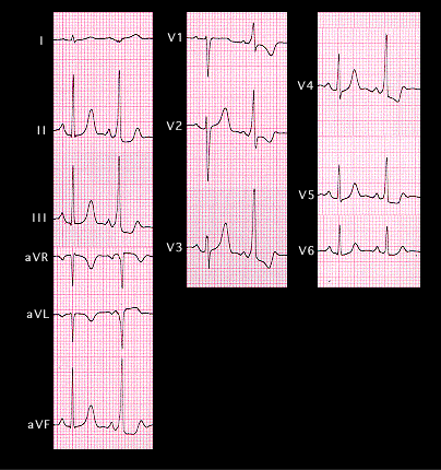
This ECG demonstrates alternating pre-excitation. The first beat of the pair is normally conducted with a normal ECG. The second is pre-excited with a delta wave that is negative in leads 1 and aVL and positive in leads V1 and V2. In addition, the R wave amplitude is greater than that of the S wave in these leads. This localizes the bypass tract to the left lateral position. Note also that the T wave is upright in leads V1,V2 and V3 in the normally conducted beat, but inverted in the pre-excited beat. These repolarization changes are secondary to the changes in the sequence of depolarization associated with the pre-excitation.