Home
Understanding the Electrocardiogram

Main navigation

  • Home
  • Understanding ECGs
    • Introduction
User account menu
  • Log in

Breadcrumb

  1. Home
  2. Understanding the Electrocardiogram In Health & Disease
  3. 3.0.0 Inter and intra-ventricular Conduction Disturbances
  4. 3.5.0 Ventricular Pre-excitation (77)

3.5.8.(85)

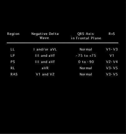
[WILL HAVE TO BE REDONE BUT NOT YET SURE HOW] The location of the bypass tract can be predicted with reasonable accuracy from the charactristics of the delta wave and the QRS complex and several somewhat complicated schemes having multiple steps have been published (see Arruda et al J Cardiovasc Electrophysiol. 9:2, 1998) A more simplified approach is shown here. It stresses the leads with negative and positive delta waves and unique charactristics of the QRS complex 1) If the bypass tract is located in the left lateral position, the delta wave will be negative or isoelectric in leads 1, aVL and V6 and positive in leads V1 and V2. In addition, the R wave amplitude will usually exceed that of the S wave in leads V1 and V2 2) If the bypass tract is located posteriorly; either left posterior, or paraseptally, the delta wave will be negative in leads II, III and aVF and positive in leads 1, aVL, V5 and V6 3) If the bypass tract is located in the right lateral position, the delta wave will be negative in lead aVR and positive in leads I, II, aVL, V5 and V6. In addition, the QRS complex will resemble left bundle branch block.

Book traversal links for 3.5.8.(85)

  • 3.5.7 (84)
  • Up
  • 3.5.9 (86)

Site is under construction

Book navigation

  • Introduction to First Edition
  • 1.0.0 Generation of the ECG
  • 2.0.0 The Normal Electrocardiogram
  • 3.0.0 Inter and intra-ventricular Conduction Disturbances
    • 3.0.1 Introduction Continued
    • 3.1.0 Right Bundle Branch Block (RBBB)
    • 3.2.0 Left Bundle Branch Block (LBBB)
    • 3.3.0 Fascicular blocks (frame 29)
    • 3.4.0 Aberrant Ventricular Conduction (52)
    • 3.5.0 Ventricular Pre-excitation (77)
      • 3.5.1
      • 3.5.2 (79)
      • 3.5.3 (80)
      • 3.5.4 (81)
      • 3.5.5 (82)
      • 3.5.6 (83)
      • 3.5.7 (84)
      • 3.5.8.(85)
      • 3.5.9 (86)
      • 3.5.10 (87)
      • 3.5.11 (88)
      • 3.5.12 ( will need new graphic)
      • 3.5.13 (89)
      • 3.5.14 (90)
      • 3.5.15 (frame 91)-last one of this chapter)
  • 4.0.0 Ventricular Hypertrophy
  • 5.0.0 ELECTROLYTE ABNORMALITIES, DRUG EFFECTS AND THE LONG QT SYNDROMES
  • 6.0.0 Ischemia and Infarction - Introduction (frame i and ii)
  • 7.0.0 Tachycardias- Introduction
  • 8.0.0 The Bradycardias frame i-introduction
  • 9.0.0 The ECG of Heart Murmurs-introduction
  • 10.0.0 The Electrocardiogram in the Emergency Department-Introduction
Powered by Drupal