Home
Understanding the Electrocardiogram

Main navigation

  • Home
  • Understanding ECGs
    • Introduction
User account menu
  • Log in

Breadcrumb

  1. Home
  2. Understanding the Electrocardiogram In Health & Disease
  3. 3.0.0 Inter and intra-ventricular Conduction Disturbances
  4. 3.5.0 Ventricular Pre-excitation (77)

3.5.10 (87)

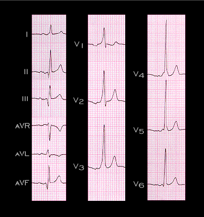
The delta wave in this tracing is negative in leads II,III and aVF, and positive in leads V1-V3. The bypass tract was in located posteriorly and on the left. The negative delta waves in leads II, III and aVF simulate the Q waves of a prior inferior wall myocardial infarction. This is referred to as a “pseudo-infarction pattern”, and the reader must be careful not to misinterpret the tracing.

Book traversal links for 3.5.10 (87)

  • 3.5.9 (86)
  • Up
  • 3.5.11 (88)

Site is under construction

Book navigation

  • Introduction to First Edition
  • 1.0.0 Generation of the ECG
  • 2.0.0 The Normal Electrocardiogram
  • 3.0.0 Inter and intra-ventricular Conduction Disturbances
    • 3.0.1 Introduction Continued
    • 3.1.0 Right Bundle Branch Block (RBBB)
    • 3.2.0 Left Bundle Branch Block (LBBB)
    • 3.3.0 Fascicular blocks (frame 29)
    • 3.4.0 Aberrant Ventricular Conduction (52)
    • 3.5.0 Ventricular Pre-excitation (77)
      • 3.5.1
      • 3.5.2 (79)
      • 3.5.3 (80)
      • 3.5.4 (81)
      • 3.5.5 (82)
      • 3.5.6 (83)
      • 3.5.7 (84)
      • 3.5.8.(85)
      • 3.5.9 (86)
      • 3.5.10 (87)
      • 3.5.11 (88)
      • 3.5.12 ( will need new graphic)
      • 3.5.13 (89)
      • 3.5.14 (90)
      • 3.5.15 (frame 91)-last one of this chapter)
  • 4.0.0 Ventricular Hypertrophy
  • 5.0.0 ELECTROLYTE ABNORMALITIES, DRUG EFFECTS AND THE LONG QT SYNDROMES
  • 6.0.0 Ischemia and Infarction - Introduction (frame i and ii)
  • 7.0.0 Tachycardias- Introduction
  • 8.0.0 The Bradycardias frame i-introduction
  • 9.0.0 The ECG of Heart Murmurs-introduction
  • 10.0.0 The Electrocardiogram in the Emergency Department-Introduction
Powered by Drupal