Home
Understanding the Electrocardiogram

Main navigation

  • Home
  • Understanding ECGs
    • Introduction
User account menu
  • Log in

Breadcrumb

  1. Home
  2. Understanding the Electrocardiogram In Health & Disease
  3. 3.0.0 Inter and intra-ventricular Conduction Disturbances
  4. 3.5.0 Ventricular Pre-excitation (77)

3.5.7 (84)

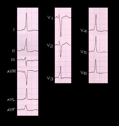
The PR interval will be shorter when the bypass tract is located in a right lateral position than when it is in a left lateral position because the distance and time required for the impulse to travel from the sinus node to the bypass tract will be shorter. This is illustrated in this ECG. The delta wave begins before the P wave is completely inscribed and the PR interval is only 0.06 seconds [CAN THE INDIVIDUAL LEADS BE MAGNIFIED AS DESIRED TO SHOW BETTER THE PR INTERVAL?] In addition, when the bypass tract is on the right, the impulse will be conducted from right to left and, as shown here, will cause a negative delta wave in lead aVR, the right sided lead, a positive delta wave in leads I, II, aVL and leads V5 and V6, the left sided leads, and a QRS complex that resembles left bundle branch block.

Book traversal links for 3.5.7 (84)

  • 3.5.6 (83)
  • Up
  • 3.5.8.(85)

Site is under construction

Book navigation

  • Introduction to First Edition
  • 1.0.0 Generation of the ECG
  • 2.0.0 The Normal Electrocardiogram
  • 3.0.0 Inter and intra-ventricular Conduction Disturbances
    • 3.0.1 Introduction Continued
    • 3.1.0 Right Bundle Branch Block (RBBB)
    • 3.2.0 Left Bundle Branch Block (LBBB)
    • 3.3.0 Fascicular blocks (frame 29)
    • 3.4.0 Aberrant Ventricular Conduction (52)
    • 3.5.0 Ventricular Pre-excitation (77)
      • 3.5.1
      • 3.5.2 (79)
      • 3.5.3 (80)
      • 3.5.4 (81)
      • 3.5.5 (82)
      • 3.5.6 (83)
      • 3.5.7 (84)
      • 3.5.8.(85)
      • 3.5.9 (86)
      • 3.5.10 (87)
      • 3.5.11 (88)
      • 3.5.12 ( will need new graphic)
      • 3.5.13 (89)
      • 3.5.14 (90)
      • 3.5.15 (frame 91)-last one of this chapter)
  • 4.0.0 Ventricular Hypertrophy
  • 5.0.0 ELECTROLYTE ABNORMALITIES, DRUG EFFECTS AND THE LONG QT SYNDROMES
  • 6.0.0 Ischemia and Infarction - Introduction (frame i and ii)
  • 7.0.0 Tachycardias- Introduction
  • 8.0.0 The Bradycardias frame i-introduction
  • 9.0.0 The ECG of Heart Murmurs-introduction
  • 10.0.0 The Electrocardiogram in the Emergency Department-Introduction
Powered by Drupal