
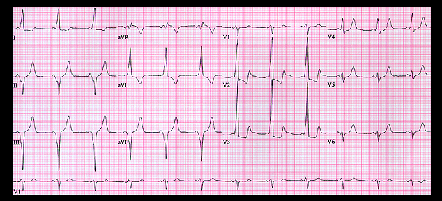
This is the ECG from the same patient recorded shortly after electrical cardioversion. It shows sinus rhythm with a PR interval of 0.08 seconds and ventricular pre-excitation. The delta wave begins before the end of the P wave and is positive in leads I, aVL, V1 and V2 and negative in leads II, III and aVF. This simulates the ECG changes of an inferior wall infarction and is referred to as a pseudo-infarction pattern. The delta wave configuration localizes the bypass tract to the right posterior paraseptal region. The QRS waveforms in this tracing and in the tracing recorded during the atrial fibrillation (displayed on the previous page) are similar. This indicates that AV conduction during atrial fibrillation was indeed via the AV nodal bypass tract and provides evidence that the refractory period of the bypass tract was shorter than that of the AV node itself.