
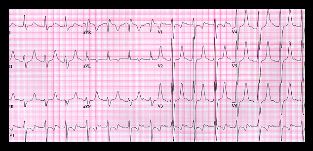
Her ECG is shown again here. It, like that of the previous patient, shows a QRS complex that is diffusely widened, measuring 0.18 seconds. P waves are present but again are somewhat difficult to identify because they are of low amplitude and are superimposed on the end of the T waves. The PR interval is prolonged (0.26 seconds), there is left axis deviation (- 30 degrees) and the T waves are peaked and symmetrical. All of these findings suggest hyperpotassemia that is somewhat less extreme than in the previous patient. In addition, the QT interval is prolonged, measuring 500 msec, and, when corrected for the rate of 83 (RR interval = 0.72 sec), is even longer (580 msec). This suggests hypocalcemia as well as hyperpotassemia and at the time of this tracing, her serum K+ was 7.2 mM and serum Ca++ was 6.3 mg/dl. This ECG, with changes that predict the combination of hyperpotassemia and hypocalcemia, is virtually diagnostic of chronic renal failure.