Home
Understanding the Electrocardiogram

Main navigation

  • Home
  • Understanding ECGs
    • Introduction
User account menu
  • Log in

Breadcrumb

  1. Home
  2. Understanding the Electrocardiogram In Health & Disease
  3. 10.0.0 The Electrocardiogram in the Emergency Department-Introduction
  4. 10.1.0 Chest pain (1)

10.1.16 (21-again)

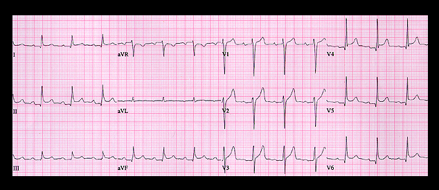
The ECG shows elevation of the ST segment in all leads except aVR. where it is depressed ,and aVL, where it is flat. Note also that in leads II and III, the PR segment is depressed relative to the TP segment. These ECG changes are highly suggestive of acute pericarditis and the physical examination revealed a pericardial friction rub, the result of uremic pericarditis.

Book traversal links for 10.1.16 (21-again)

  • 10.1.15 (frame 21).
  • Up
  • 10.2.0 Shortness of breath (frame 23)

Site is under construction

Book navigation

  • Introduction to First Edition
  • 1.0.0 Generation of the ECG
  • 2.0.0 The Normal Electrocardiogram
  • 3.0.0 Inter and intra-ventricular Conduction Disturbances
  • 4.0.0 Ventricular Hypertrophy
  • 5.0.0 ELECTROLYTE ABNORMALITIES, DRUG EFFECTS AND THE LONG QT SYNDROMES
  • 6.0.0 Ischemia and Infarction - Introduction (frame i and ii)
  • 7.0.0 Tachycardias- Introduction
  • 8.0.0 The Bradycardias frame i-introduction
  • 9.0.0 The ECG of Heart Murmurs-introduction
  • 10.0.0 The Electrocardiogram in the Emergency Department-Introduction
    • 10.1.0 Chest pain (1)
      • 10.1.1 (1-again)
      • 10.1.2(2)
      • 10.1.3 (frame 4)
      • 10.1.4 (5))
      • 10.1.5 (7)
      • 10.1.6 (8)
      • 10.1.7 (10)
      • 10.1.8 (10-again)
      • 10.1.9 (12)
      • 10.1.10(13)
      • 10.1.11 (14)
      • 10.1.12 (15)
      • 10.1.13 (15 again)
      • 10.1.14 (17)
      • 10.1.15 (frame 21).
      • 10.1.16 (21-again)
    • 10.2.0 Shortness of breath (frame 23)
    • 10.3.0 Palpitations (frame 31)
    • 10.4.0 confusion, weakness etc (frame 40)
    • 10.5.0 Conclusion (no image)
Powered by Drupal