
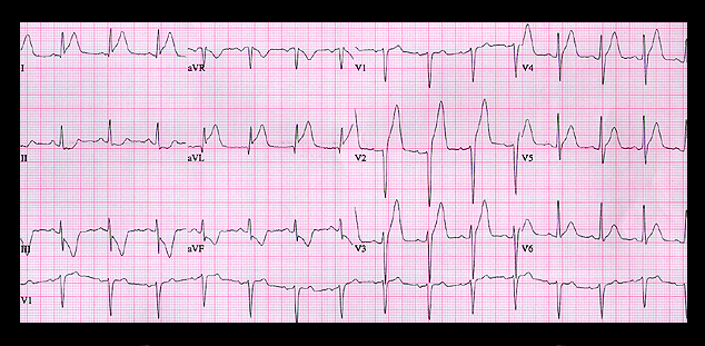
The ECG shows rather obvious changes of an acute anterior wall myocardial infarction with ST segment elevation and peaked T waves in leads I, aVL and V2-V6. ST segment depression and inverted T waves are seen in leads III and aVF. These are the reciprocal of the ST segment and T wave changes in leads I and aVL. These ECG changes predict that the site of the occlusion is in the proximal portion of left anterior descending coronary artery, above the 1st septal perforator. An emergency cardiac catheterization showed a totally occluded left anterior descending coronary artery at the predicted location and this was successfully angioplastied.