Home
Understanding the Electrocardiogram

Main navigation

  • Home
  • Understanding ECGs
    • Introduction
User account menu
  • Log in

Breadcrumb

  1. Home
  2. Understanding the Electrocardiogram In Health & Disease
  3. 10.0.0 The Electrocardiogram in the Emergency Department-Introduction
  4. 10.1.0 Chest pain (1)

10.1.9 (12)

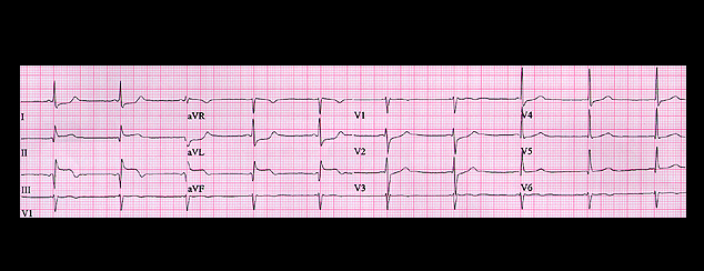
The tracing shown here is from a 54 year old female who presented to the Emergency Department with chest pain and a blood pressure of 90/60. Her ECG shows rather obvious changes of an acute inferior wall infarction. But what else does it suggest? What is the rhythm and where would you place the lesion causing the infarction?

Book traversal links for 10.1.9 (12)

  • 10.1.8 (10-again)
  • Up
  • 10.1.10(13)

Site is under construction

Book navigation

  • Introduction to First Edition
  • 1.0.0 Generation of the ECG
  • 2.0.0 The Normal Electrocardiogram
  • 3.0.0 Inter and intra-ventricular Conduction Disturbances
  • 4.0.0 Ventricular Hypertrophy
  • 5.0.0 ELECTROLYTE ABNORMALITIES, DRUG EFFECTS AND THE LONG QT SYNDROMES
  • 6.0.0 Ischemia and Infarction - Introduction (frame i and ii)
  • 7.0.0 Tachycardias- Introduction
  • 8.0.0 The Bradycardias frame i-introduction
  • 9.0.0 The ECG of Heart Murmurs-introduction
  • 10.0.0 The Electrocardiogram in the Emergency Department-Introduction
    • 10.1.0 Chest pain (1)
      • 10.1.1 (1-again)
      • 10.1.2(2)
      • 10.1.3 (frame 4)
      • 10.1.4 (5))
      • 10.1.5 (7)
      • 10.1.6 (8)
      • 10.1.7 (10)
      • 10.1.8 (10-again)
      • 10.1.9 (12)
      • 10.1.10(13)
      • 10.1.11 (14)
      • 10.1.12 (15)
      • 10.1.13 (15 again)
      • 10.1.14 (17)
      • 10.1.15 (frame 21).
      • 10.1.16 (21-again)
    • 10.2.0 Shortness of breath (frame 23)
    • 10.3.0 Palpitations (frame 31)
    • 10.4.0 confusion, weakness etc (frame 40)
    • 10.5.0 Conclusion (no image)
Powered by Drupal