
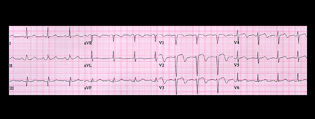
This is the ECG recorded on the next day. Note the decrease in R wave amplitude and the increase in S wave amplitude in leads V1-V5. These changes are probably due to changes in the placement of the chest leads, but could also be due to an infarction. The T waves are now symmetrically inverted in leads V2-V4. These T wave changes are consistent with an evolving ischemic changes and are sometimes referred to as "post ischemic T-wave changes". Her cardiac enzymes were elevated, and a coronary angiogram, performed after transfer to a center with cardiac catheterization capabilities, revealed a complete occlusion in the mid-portion of the anterior descending coronary artery .