
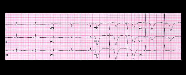
This ECG, from the same patient, was recorded 12 hours later. The T waves are more deeply inverted and the QT interval is more prolonged. T waves of this type are referred to as “Giant T Waves” and when they occur in a patient with an intracranial hemorrhage, they are referred to as the “CVA Pattern”. In our patient they were caused by the acute ischemic event and the coronary angiogram revealed a 95% occlusion in the proximal portion of the left anterior descending coronary artery.