Home
Understanding the Electrocardiogram

Main navigation

  • Home
  • Understanding ECGs
    • Introduction
User account menu
  • Log in

Breadcrumb

  1. Home
  2. Understanding the Electrocardiogram In Health & Disease
  3. 7.0.0 Tachycardias- Introduction
  4. 7.4.0 Wide Complex Beats and Rhythms (frame 122)
  5. 7.4.30 (frame 154)

7.4.36 (160)

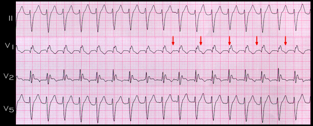
The RR interval is 0.40 seconds (rate 150) and the QRS duration is 0.13 seconds. The QRS configuration is quite typical for right bundle branch block with a frontal plane axis of -76 degrees, an indication of left anterior fascicular block in addition to the right bundle branch bloc . There are RS complexes in the precordial leads and the interval from the onset of the R wave to the nadir of the S wave is 60 ms. These features are consistent with a diagnosis of supraventricular tachycardia conducted with aberration.

It is difficult to identify P waves at first glance, but on closer inspection, there are subtle deflections in the ST segment, seen best in lead V1 (arrows) that are consistent with P waves and are dissociated from the QRS complexes. Their rate is 90 per minute. The finding of AV dissociation establishes this tachycardia as being ventricular in origin.

The QRS waveform suggests that the origin of the tachycardia is in the posterior fascicle of the left bundle. This rhythm is referred to as a fascicular tachycardia. It is a common form of the idiopathic ventricular tachycardias that occur primarily in younger patients with no evidence of underlying heart disease, and it usually responds to treatment with calcium channel blocking agents.

Book traversal links for 7.4.36 (160)

  • 7.4.35 (159)
  • Up
  • 7.4.37 (161)

Site is under construction

Book navigation

  • Introduction to First Edition
  • 1.0.0 Generation of the ECG
  • 2.0.0 The Normal Electrocardiogram
  • 3.0.0 Inter and intra-ventricular Conduction Disturbances
  • 4.0.0 Ventricular Hypertrophy
  • 5.0.0 ELECTROLYTE ABNORMALITIES, DRUG EFFECTS AND THE LONG QT SYNDROMES
  • 6.0.0 Ischemia and Infarction - Introduction (frame i and ii)
  • 7.0.0 Tachycardias- Introduction
    • 7.1.0 Mechanisms of arrhythmias-frame 1
    • 7.2.0 Narrow Complex Rhythms (frame 38)
    • 7.3.0 Atrial Flutter and Fibrillation (frame 93)
    • 7.4.0 Wide Complex Beats and Rhythms (frame 122)
      • 7.4.1 (123)
      • 7.4.2 (124)
      • 7.4.3 (125)
      • 7.4.4 (126)
      • 7.4.5 (127)
      • 7.4.6 (128)
      • 7.4.7 (129)
      • 7.4.8 (130)
      • 7.4.9 (131)
      • 7.4.10 (frame 132)
      • 7.4.20 (frame 142)
      • 7.4.30 (frame 154)
        • 7.4.31 (155)
        • 7.4.32 (156)
        • 7.4.33 (157)
        • 7.4.34 (158)
        • 7.4.35 (159)
        • 7.4.36 (160)
        • 7.4.37 (161)
        • 7.4.38 (161-again)
        • 7.4.39 (162)
      • 7.4.40 (frame 163)
  • 8.0.0 The Bradycardias frame i-introduction
  • 9.0.0 The ECG of Heart Murmurs-introduction
  • 10.0.0 The Electrocardiogram in the Emergency Department-Introduction
Powered by Drupal