Home
Understanding the Electrocardiogram

Main navigation

  • Home
  • Understanding ECGs
    • Introduction
User account menu
  • Log in

Breadcrumb

  1. Home
  2. Understanding the Electrocardiogram In Health & Disease
  3. 7.0.0 Tachycardias- Introduction
  4. 7.4.0 Wide Complex Beats and Rhythms (frame 122)
  5. 7.4.30 (frame 154)

7.4.32 (156)

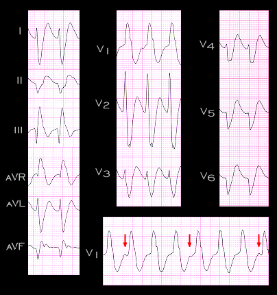
Another example demonstrating the usefulness of these various criteria is shown here. This wide complex tachycardia has an RR interval of 0.44 seconds (rate 136). The tracing is from a 60 year old male who also had a history of a prior myocardial infarction. The QRS complex measures 0.18 seconds and suggests right bundle branch block. The QRS complex in V1 shows a single positive component and the interval from the onset of the R wave to the nadir of the S wave in leads V2 and V3 exceeds 100ms. In addition, P waves can be identified in lead V1 (arrows) that are dissociated from the QRS complexes. These features identify this wide complex tachycardia as being ventricular in origin.

Book traversal links for 7.4.32 (156)

  • 7.4.31 (155)
  • Up
  • 7.4.33 (157)

Site is under construction

Book navigation

  • Introduction to First Edition
  • 1.0.0 Generation of the ECG
  • 2.0.0 The Normal Electrocardiogram
  • 3.0.0 Inter and intra-ventricular Conduction Disturbances
  • 4.0.0 Ventricular Hypertrophy
  • 5.0.0 ELECTROLYTE ABNORMALITIES, DRUG EFFECTS AND THE LONG QT SYNDROMES
  • 6.0.0 Ischemia and Infarction - Introduction (frame i and ii)
  • 7.0.0 Tachycardias- Introduction
    • 7.1.0 Mechanisms of arrhythmias-frame 1
    • 7.2.0 Narrow Complex Rhythms (frame 38)
    • 7.3.0 Atrial Flutter and Fibrillation (frame 93)
    • 7.4.0 Wide Complex Beats and Rhythms (frame 122)
      • 7.4.1 (123)
      • 7.4.2 (124)
      • 7.4.3 (125)
      • 7.4.4 (126)
      • 7.4.5 (127)
      • 7.4.6 (128)
      • 7.4.7 (129)
      • 7.4.8 (130)
      • 7.4.9 (131)
      • 7.4.10 (frame 132)
      • 7.4.20 (frame 142)
      • 7.4.30 (frame 154)
        • 7.4.31 (155)
        • 7.4.32 (156)
        • 7.4.33 (157)
        • 7.4.34 (158)
        • 7.4.35 (159)
        • 7.4.36 (160)
        • 7.4.37 (161)
        • 7.4.38 (161-again)
        • 7.4.39 (162)
      • 7.4.40 (frame 163)
  • 8.0.0 The Bradycardias frame i-introduction
  • 9.0.0 The ECG of Heart Murmurs-introduction
  • 10.0.0 The Electrocardiogram in the Emergency Department-Introduction
Powered by Drupal