
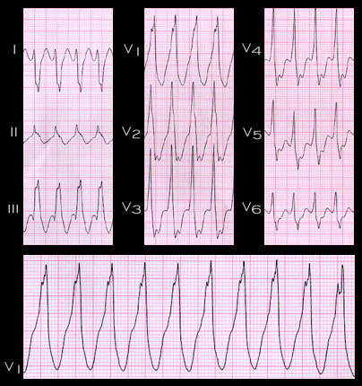
The ECG shown here reveals a wide complex tachycardia with an RR interval of 0.36 seconds (rate 165). It is from a 56 year old male with a history of a prior anterior wall myocardial infarction. The QRS complex measures 0.166 seconds (166 msec) and shows right bundle branch block with right axis deviation (+127 degrees). AV dissociation can not be detected. The patient’s age, and past history, and a QRS duration greater than 0.14 seconds are more consistent with a diagnosis of ventricular tachycardia than of supraventricular tachycardia with aberrancy, as is the QRS morphology in V1. In addition, the interval from the onset of the R wave to the nadir of the S wave in leads V3 to V6 exceeds 100ms. This is highly suggestive of ventricular tachycardia, since in the study of Brugada et al, 172 of 175 patients fulfilling this criterion were found to have ventricular tachycardia.