Home
Understanding the Electrocardiogram

Main navigation

  • Home
  • Understanding ECGs
    • Introduction
User account menu
  • Log in

Breadcrumb

  1. Home
  2. Understanding the Electrocardiogram In Health & Disease
  3. 7.0.0 Tachycardias- Introduction
  4. 7.2.0 Narrow Complex Rhythms (frame 38)
  5. 7.2.10 (frame 48)

7.2.14 (52)

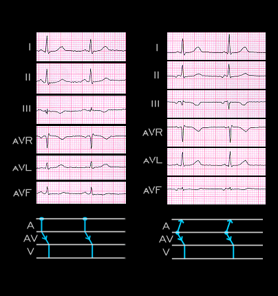
The sinus P wave from page 7.2.2 is shown on the left. Its frontal plane vector is directed to the left and inferiorly with an axis of +60 degrees. The abnormal P wave from the previous page is shown on the right.

Its frontal plane axis is still ponted to the left but it is now directed superiorly reflecting the superiorly directed activation of the atria. Its axis is -70 degrees. The PR interval of the sinus P wave is 0.16 seconds while that associated with the ectopic P wave is 0.10 seconds. This suggests that the ectopic focus is located within, or very close to the AV node, above the region of the AV nodal delay, and that it activates the atria in a retrograde direction before traveling through the rest of the AV node and His-Purkinje system to activate the ventricles. The ladder diagrams shown below the ECGs are a graphic way to illustrate these relationships.

Book traversal links for 7.2.14 (52)

  • 7.2.13 (51)
  • Up
  • 7.2.15 (53)

Site is under construction

Book navigation

  • Introduction to First Edition
  • 1.0.0 Generation of the ECG
  • 2.0.0 The Normal Electrocardiogram
  • 3.0.0 Inter and intra-ventricular Conduction Disturbances
  • 4.0.0 Ventricular Hypertrophy
  • 5.0.0 ELECTROLYTE ABNORMALITIES, DRUG EFFECTS AND THE LONG QT SYNDROMES
  • 6.0.0 Ischemia and Infarction - Introduction (frame i and ii)
  • 7.0.0 Tachycardias- Introduction
    • 7.1.0 Mechanisms of arrhythmias-frame 1
    • 7.2.0 Narrow Complex Rhythms (frame 38)
      • 7.2.1 (39)
      • 7.2.2 (FRAME 40)
      • 7.2.3 (41)
      • 7.2.4 (42)
      • 7.2.5 (43)
      • 7.2.6 (44)
      • 7.2.7 (45)
      • 7.2.8 (46)
      • 7.2.9 (47)
      • 7.2.10 (frame 48)
        • 7.2.11 (49)
        • 7.2.12 (50)
        • 7.2.13 (51)
        • 7.2.14 (52)
        • 7.2.15 (53)
        • 7.2.16 (54)
        • 7.2.17 (55)
        • 7.2.18 (56)
        • 7.2.19 (57)
      • 7.2.20 (frame 58)
      • 7.2.30 (frame 68)
      • 7.2.40 (frame 78)
      • 7.2.50 (frame 88)
    • 7.3.0 Atrial Flutter and Fibrillation (frame 93)
    • 7.4.0 Wide Complex Beats and Rhythms (frame 122)
  • 8.0.0 The Bradycardias frame i-introduction
  • 9.0.0 The ECG of Heart Murmurs-introduction
  • 10.0.0 The Electrocardiogram in the Emergency Department-Introduction
Powered by Drupal