Home
Understanding the Electrocardiogram

Main navigation

  • Home
  • Understanding ECGs
    • Introduction
User account menu
  • Log in

Breadcrumb

  1. Home
  2. Understanding the Electrocardiogram In Health & Disease
  3. 7.0.0 Tachycardias- Introduction
  4. 7.2.0 Narrow Complex Rhythms (frame 38)

7.2.8 (46)

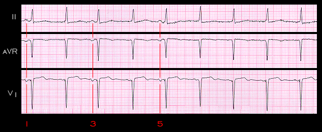
An ectopic atrial pacemaker located in close proximity to the sinus node will depolarize the atria in a sequence that is nearly normal. For this reason, the ectopic P wave will appear nearly normal. If the sinus rate and the ectopic rate are similar, the ECG will suggest a wandering atrial pacemaker. The ECG shown here is from a 78 year old male patient with pulmonary edema. The morphology of the P wave changes slightly, as in the previous example, but the P-P interval does not change in a phasic fashion. This suggests that the P wave changes most likely reflect an ectopic focus whose rate is similar to that of the sinus node. The observation that the two P wave morphologies are quite similar suggests that the ectopic focus is located in close proximity to the sinus node. When, as in this case, the sinus and ectopic rates are similar, they are said to be “isorhythmic”. The P waves labeled 1, 3, and 5 are the sinus P waves. The other P waves are ectopic.

Book traversal links for 7.2.8 (46)

  • 7.2.7 (45)
  • Up
  • 7.2.9 (47)

Site is under construction

Book navigation

  • Introduction to First Edition
  • 1.0.0 Generation of the ECG
  • 2.0.0 The Normal Electrocardiogram
  • 3.0.0 Inter and intra-ventricular Conduction Disturbances
  • 4.0.0 Ventricular Hypertrophy
  • 5.0.0 ELECTROLYTE ABNORMALITIES, DRUG EFFECTS AND THE LONG QT SYNDROMES
  • 6.0.0 Ischemia and Infarction - Introduction (frame i and ii)
  • 7.0.0 Tachycardias- Introduction
    • 7.1.0 Mechanisms of arrhythmias-frame 1
    • 7.2.0 Narrow Complex Rhythms (frame 38)
      • 7.2.1 (39)
      • 7.2.2 (FRAME 40)
      • 7.2.3 (41)
      • 7.2.4 (42)
      • 7.2.5 (43)
      • 7.2.6 (44)
      • 7.2.7 (45)
      • 7.2.8 (46)
      • 7.2.9 (47)
      • 7.2.10 (frame 48)
      • 7.2.20 (frame 58)
      • 7.2.30 (frame 68)
      • 7.2.40 (frame 78)
      • 7.2.50 (frame 88)
    • 7.3.0 Atrial Flutter and Fibrillation (frame 93)
    • 7.4.0 Wide Complex Beats and Rhythms (frame 122)
  • 8.0.0 The Bradycardias frame i-introduction
  • 9.0.0 The ECG of Heart Murmurs-introduction
  • 10.0.0 The Electrocardiogram in the Emergency Department-Introduction
Powered by Drupal