Home
Understanding the Electrocardiogram

Main navigation

  • Home
  • Understanding ECGs
    • Introduction
User account menu
  • Log in

Breadcrumb

  1. Home
  2. Understanding the Electrocardiogram In Health & Disease
  3. 7.0.0 Tachycardias- Introduction
  4. 7.2.0 Narrow Complex Rhythms (frame 38)

7.2.50 (frame 88)

In approximately 5-10% of cases, the location of the antegrade and retrograde pathways are reversed The fast pathway becomes the antegrade limb of the re-entry circuit and the slow pathway, the retrograde limb. When this occurs, the PR interval becomes the shorter interval and the RP interval becomes the longer interval. In AV nodal reentrant tachycardias (AVNRT), where the pathways are in or close to the AV node, the shift in pathways will affect the PR and RP intervals, but not the QRS complex. In AVRT, in which one pathway is a bypass tract, antegrade conduction over this, the rapidly conducting pathway pathway, and retrograde conduction via the AV node, will affect the QRS complex as well as the PR and RP intervals.

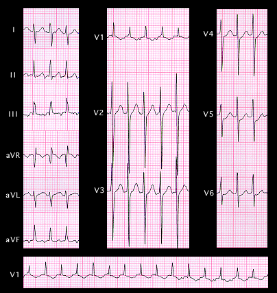
This ECG reveals a narrow complex tachycardia with a rate of 220 beats per minute. There are P waves before each QRS complex, occurring coincident with the end of the T wave. They are inverted in leads II, III and aVF and upright in leads aVR and aVL. The PR interval is 0.10 seconds and the interval from the onset of the QRS complex to the following P wave (the RP interval) is 0.16 seconds. This ECG could be interpreted as either a rapid ectopic atrial tachycardia or as an AV nodal reentrant tachycardia (AVNRT) in which the antegrade conduction occurs over the fast pathway, and retrograde conduction is via the slow pathway, thereby accounting for the short PR interval and the longer RP interval

  • 7.2.51 (89)
  • 7.2.52 (90)
  • 7.2.53 (91}
  • 7.2.54 (92)

Book traversal links for 7.2.50 (frame 88)

  • 7.2.49 (87)
  • Up
  • 7.2.51 (89)

Site is under construction

Book navigation

  • Introduction to First Edition
  • 1.0.0 Generation of the ECG
  • 2.0.0 The Normal Electrocardiogram
  • 3.0.0 Inter and intra-ventricular Conduction Disturbances
  • 4.0.0 Ventricular Hypertrophy
  • 5.0.0 ELECTROLYTE ABNORMALITIES, DRUG EFFECTS AND THE LONG QT SYNDROMES
  • 6.0.0 Ischemia and Infarction - Introduction (frame i and ii)
  • 7.0.0 Tachycardias- Introduction
    • 7.1.0 Mechanisms of arrhythmias-frame 1
    • 7.2.0 Narrow Complex Rhythms (frame 38)
      • 7.2.1 (39)
      • 7.2.2 (FRAME 40)
      • 7.2.3 (41)
      • 7.2.4 (42)
      • 7.2.5 (43)
      • 7.2.6 (44)
      • 7.2.7 (45)
      • 7.2.8 (46)
      • 7.2.9 (47)
      • 7.2.10 (frame 48)
      • 7.2.20 (frame 58)
      • 7.2.30 (frame 68)
      • 7.2.40 (frame 78)
      • 7.2.50 (frame 88)
        • 7.2.51 (89)
        • 7.2.52 (90)
        • 7.2.53 (91}
        • 7.2.54 (92)
    • 7.3.0 Atrial Flutter and Fibrillation (frame 93)
    • 7.4.0 Wide Complex Beats and Rhythms (frame 122)
  • 8.0.0 The Bradycardias frame i-introduction
  • 9.0.0 The ECG of Heart Murmurs-introduction
  • 10.0.0 The Electrocardiogram in the Emergency Department-Introduction
Powered by Drupal