
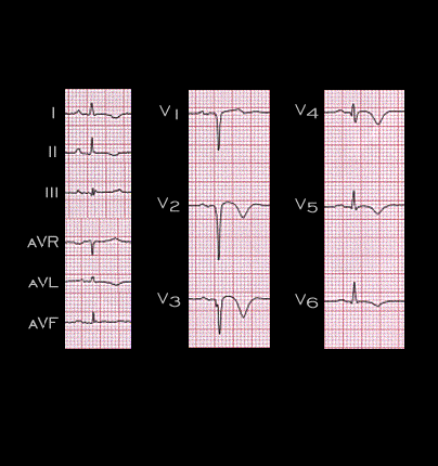
The third type of T wave changes, the chronic changes, characteristically occur after the changes associated with the acute ischemic event have evolved. This ECG is from the 74 year old female whose ECGs, recorded during an acute anterior wall infarction, were shown earlier (see pages 6.1.6 and 6.2.4). This ECG was recorded one year after the acute event and demonstrates persistently inverted T waves in all chest leads and in leads I, II and aVL. Note also the QS complex in leads V1-V3 and the small Q wave in lead V4 which indicate the transmural loss of muscle mass in the anterior wall of the left ventricle.