Home
Understanding the Electrocardiogram

Main navigation

  • Home
  • Understanding ECGs
    • Introduction
User account menu
  • Log in

Breadcrumb

  1. Home
  2. Understanding the Electrocardiogram In Health & Disease
  3. 5.0.0 ELECTROLYTE ABNORMALITIES, DRUG EFFECTS AND THE LONG QT SYNDROMES
  4. 5.5.0 Drug effects (frame 45)
  5. 5.5.40 (frame 85)

5.5.49(94)

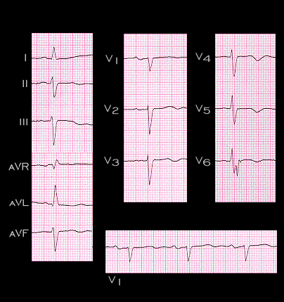
This tracing is from the same patient, recorded one day after discontinuing digitalis.  The ectopic atrial tachycardia and AV block indicative of digitalis toxicity are no longer present. The atrial rate is now 60/min and every P wave is conducted to the ventricles with a PR interval that is only slightly prolonged (0.22 seconds). The P wave morphology is now normal and the P wave axis is +30 degrees, indicating that the P waves are originating in (or very close to) the sinus node.

Book traversal links for 5.5.49(94)

  • 5.5.48 (93)
  • Up
  • 5.5.50 (frame 95)

Site is under construction

Book navigation

  • Introduction to First Edition
  • 1.0.0 Generation of the ECG
  • 2.0.0 The Normal Electrocardiogram
  • 3.0.0 Inter and intra-ventricular Conduction Disturbances
  • 4.0.0 Ventricular Hypertrophy
  • 5.0.0 ELECTROLYTE ABNORMALITIES, DRUG EFFECTS AND THE LONG QT SYNDROMES
    • 5.1.0 Ion Channel R review (frame 1)
    • 5.2.0 High Potassium (9)
    • 5.2.10 (frame 19)
    • 5.3.0 low potassium (frame 28)
    • 5.4.0 CHANGES IN CALCIUM AND SODIUM (frame 36)
    • 5.5.0 Drug effects (frame 45)
      • 5.5.1 (frame 46)
      • 5.5.2 (47)
      • 5.5.3 (48)
      • 5.5.4(49)
      • 5.5.5(50)
      • 5.5.6 (51)
      • 5.5.7 (52)
      • 5.5.8 (53)
      • 5.5.9 (54)
      • 5.5.10 (55)
      • 5.5.20(frame 65)
      • 5.5.30 (frame 75)
      • 5.5.40 (frame 85)
        • 5.5.41 (86)
        • 5.5.42 (87)
        • 5.5.43(88)
        • 5.5.44(89)
        • 5.5.45(90)
        • 5.5.46(91)
        • 5.5.47 (92)
        • 5.5.48 (93)
        • 5.5.49(94)
      • 5.5.50 (frame 95)
    • 5.6.0 (frame 96) Hypothermia
    • 5.7.0 Brugada syndrome frame 116
  • 6.0.0 Ischemia and Infarction - Introduction (frame i and ii)
  • 7.0.0 Tachycardias- Introduction
  • 8.0.0 The Bradycardias frame i-introduction
  • 9.0.0 The ECG of Heart Murmurs-introduction
  • 10.0.0 The Electrocardiogram in the Emergency Department-Introduction
Powered by Drupal