Home
Understanding the Electrocardiogram

Main navigation

  • Home
  • Understanding ECGs
    • Introduction
User account menu
  • Log in

Breadcrumb

  1. Home
  2. Understanding the Electrocardiogram In Health & Disease
  3. 2.0.0 The Normal Electrocardiogram
  4. 2.4.0 ST segment

2.4.1

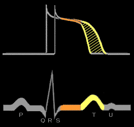
The ST segment reflects the phase of slow repolarization which is represented by the plateau phase (phase 2) of the action potential and which, under normal circumstances, is at essentially the same voltage level in all ventricular myocardial cells. For this reason, there are no voltage gradients during this portion of repolarization and the representative electrocardiographic waveform (the ST segment) is isoelectric, i.e., does not deviate from baseline. The lack of voltage gradients during this phase is similar to the lack of voltage gradients during the resting phase and explains why both the ST segment and the TP segment are isoelectric, even though they represent different transmembrane voltages. The T wave, in contrast, reflects the voltage gradients created during the phase of rapid repolarization (phase 3) of the ventricular myocardial cells as they sequentially repolarize. These two different phases of repolarization are affected differently by a large variety of physiologic, pharmacologic and pathologic factors and for this reason, should be thought of separately.

Book traversal links for 2.4.1

  • 2.4.0 ST segment
  • Up
  • 2.4.2

Site is under construction

Book navigation

  • Introduction to First Edition
  • 1.0.0 Generation of the ECG
  • 2.0.0 The Normal Electrocardiogram
    • 2.1.0 P Wave
    • 2.2.0 PR Interval
    • 2.3.0 QRS Complex
    • 2.4.0 ST segment
      • 2.4.1
      • 2.4.2
      • 2.4.3
    • 2.5.0 T wave
    • 2.6.0 QT Interval
    • 2.7.0 U Wave
  • 3.0.0 Inter and intra-ventricular Conduction Disturbances
  • 4.0.0 Ventricular Hypertrophy
  • 5.0.0 ELECTROLYTE ABNORMALITIES, DRUG EFFECTS AND THE LONG QT SYNDROMES
  • 6.0.0 Ischemia and Infarction - Introduction (frame i and ii)
  • 7.0.0 Tachycardias- Introduction
  • 8.0.0 The Bradycardias frame i-introduction
  • 9.0.0 The ECG of Heart Murmurs-introduction
  • 10.0.0 The Electrocardiogram in the Emergency Department-Introduction
Powered by Drupal