Home
Understanding the Electrocardiogram

Main navigation

  • Home
  • Understanding ECGs
    • Introduction
User account menu
  • Log in

Breadcrumb

  1. Home
  2. Understanding the Electrocardiogram In Health & Disease
  3. 9.0.0 The ECG of Heart Murmurs-introduction
  4. 9.1.0 Systolic Heart Murmurs (frame 3)
  5. 9.1.20 (29)

9.1.27 (37)

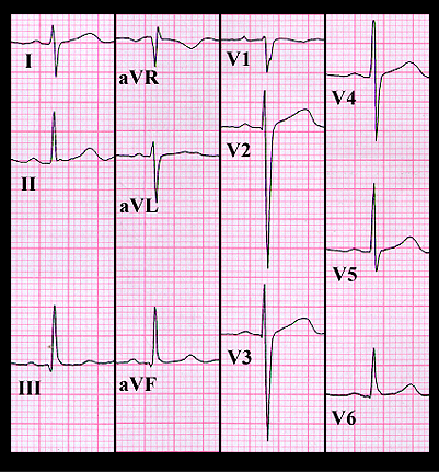
Her ECG, shown again here, reveals sinus rhythm with normal PR, QRS and QT intervals. The frontal plane QRS spatial vector is directed inferiorly and slightly to the right with an axis of +100 degrees. The amplitude of the S waves in leads V2 and V3 are at the upper limits of normal for a woman of this age. These ECG findings, in combination with the systolic click and murmur suggest mitral valve prolapse with mild mitral insufficiency. This diagnosis was confirmed by the echocardiogram.

Book traversal links for 9.1.27 (37)

  • 9.1.26 (36)
  • Up
  • 9.2.0 Diastolic Murmurs (frame 38)

Site is under construction

Book navigation

  • Introduction to First Edition
  • 1.0.0 Generation of the ECG
  • 2.0.0 The Normal Electrocardiogram
  • 3.0.0 Inter and intra-ventricular Conduction Disturbances
  • 4.0.0 Ventricular Hypertrophy
  • 5.0.0 ELECTROLYTE ABNORMALITIES, DRUG EFFECTS AND THE LONG QT SYNDROMES
  • 6.0.0 Ischemia and Infarction - Introduction (frame i and ii)
  • 7.0.0 Tachycardias- Introduction
  • 8.0.0 The Bradycardias frame i-introduction
  • 9.0.0 The ECG of Heart Murmurs-introduction
    • 9.0.1 (frame 1)
    • 9.0.2 (2)
    • 9.1.0 Systolic Heart Murmurs (frame 3)
      • 9.1.1 (4)
      • 9.1.2 (frame 4 again)
      • 9.1.3 (5)
      • 9.1.4 (6)
      • 9.1.5 (7)
      • 9.1.6 (8)
      • 9.1.7 (9)
      • 9.1.8 (10)
      • 9.1.9 (11)
      • 9.1.10 (12)
      • 9.1.20 (29)
        • 9.1.21 (30)
        • 9.1.22 (32-omit frame 31)
        • 9.1.23 (33)
        • 9.1.24 (34)
        • 9.1.25 (35)
        • 9.1.26 (36)
        • 9.1.27 (37)
    • 9.2.0 Diastolic Murmurs (frame 38)
    • 9.3.0 Systolic and Diastolic Murmurs (frame 43)
  • 10.0.0 The Electrocardiogram in the Emergency Department-Introduction
Powered by Drupal