
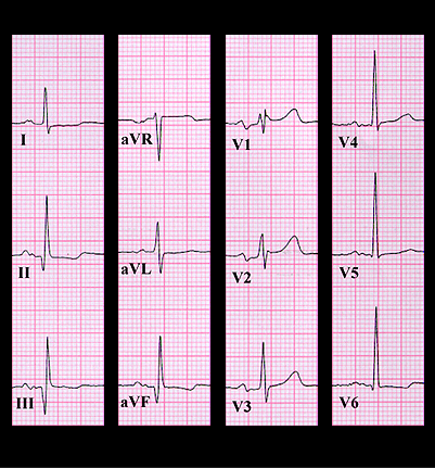
His ECG is shown again here. There is sinus rhythm and the PR interval, QRS duration and QT interval are normal. The P wave is notched in leads II, III and aVF and has a broad negative component in V1. This suggests left atrial enlargement. The Q waves in leads II, II and aVF and broad waves in leads V1 and V2 are consistent with a prior inferior wall infarction that also involved the posterior wall. The late systolic crescendo murmur is quite typical of the mitral insufficiency caused by a prolapsing mitral valve leaflet. In this case, the prolapsing leaflet was the result of papillary muscle insufficiency, a consequence of the prior inferior/posterior infarction. This diagnosis was confirmed by the echocardiogram which demonstrated prolapse of the posterior leaflet of the mitral valve in the latter half of systole causing moderate mitral insufficiency.