
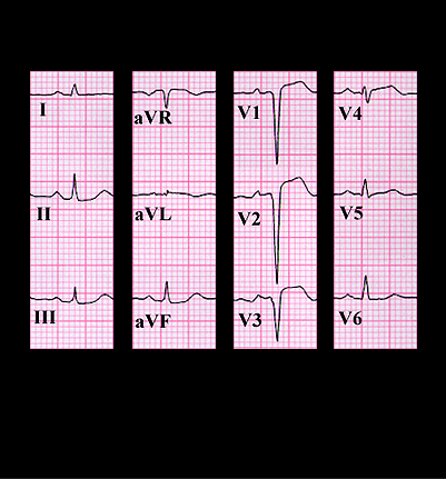
The ECG from the previous page is shown again here. Note that the ST segments are still elevated in leads V2-V4 but the T waves are less prominent. A QS complex is now present in leads V1-V3 and there is a small Q wave in V4. These are the evolving changes of an acute anterior infarction. The appearance of a new heart murmur suggests a complication of the infarction and the differential includes a ruptured papillary muscle causing acute mitral insufficiency or a perforated interventricular septum with the creation of a ventricular septal defect and a left to right shunt. A ruptured papillary muscle occurs more frequently in association with an acute inferior wall infarction whereas the perforated septum is more common in the setting of an acute anterior wall infarction and this was the cause of the murmur in this patient.