Home
Understanding the Electrocardiogram

Main navigation

  • Home
  • Understanding ECGs
    • Introduction
User account menu
  • Log in

Breadcrumb

  1. Home
  2. Understanding the Electrocardiogram In Health & Disease
  3. 7.0.0 Tachycardias- Introduction
  4. 7.2.0 Narrow Complex Rhythms (frame 38)
  5. 7.2.40 (frame 78)

7.2.45 (83)

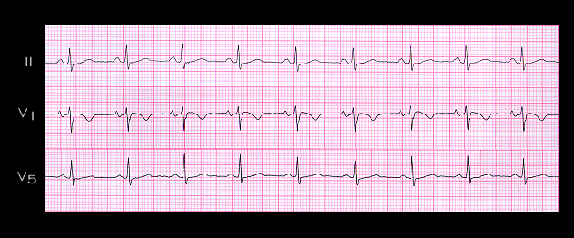
This tracing, from the same patient, was recorded shortly after the intravenous administration of adenosine. There is now sinus rhythm with a rate of 80 per minute and a PR interval of approximately 0.16 seconds. The deflection suggestive of an R’ in V1 is no longer present, confirming that it was caused by a retrograde P wave. The ST segment depression seen in leads II and V5 during the tachycardia is also no longer present. This could also have been a manifestation of the retrograde P wave, or an indication of ischemia induced by the sudden increase in rate. The presence of the retrograde P wave in or beyond the end of the QRS complex is consistent with the retrograde pathways being either within the AV node and perinodal tissue, or  in an AV nodal bypass tract. In general, the more displaced the retrograde P wave from the end of the QRS complex, the more likely it is that an AV nodal bypass tract formed the retrograde limb of the re-entry circuit

Book traversal links for 7.2.45 (83)

  • 7.2.44 (82)
  • Up
  • 7.2.46 (84)

Site is under construction

Book navigation

  • Introduction to First Edition
  • 1.0.0 Generation of the ECG
  • 2.0.0 The Normal Electrocardiogram
  • 3.0.0 Inter and intra-ventricular Conduction Disturbances
  • 4.0.0 Ventricular Hypertrophy
  • 5.0.0 ELECTROLYTE ABNORMALITIES, DRUG EFFECTS AND THE LONG QT SYNDROMES
  • 6.0.0 Ischemia and Infarction - Introduction (frame i and ii)
  • 7.0.0 Tachycardias- Introduction
    • 7.1.0 Mechanisms of arrhythmias-frame 1
    • 7.2.0 Narrow Complex Rhythms (frame 38)
      • 7.2.1 (39)
      • 7.2.2 (FRAME 40)
      • 7.2.3 (41)
      • 7.2.4 (42)
      • 7.2.5 (43)
      • 7.2.6 (44)
      • 7.2.7 (45)
      • 7.2.8 (46)
      • 7.2.9 (47)
      • 7.2.10 (frame 48)
      • 7.2.20 (frame 58)
      • 7.2.30 (frame 68)
      • 7.2.40 (frame 78)
        • 7.2.41 (79)
        • 7.2.42 (80)
        • 7.2.43 (81)
        • 7.2.44 (82)
        • 7.2.45 (83)
        • 7.2.46 (84)
        • 7.2.47 (85)
        • 7.2.48 (86)
        • 7.2.49 (87)
      • 7.2.50 (frame 88)
    • 7.3.0 Atrial Flutter and Fibrillation (frame 93)
    • 7.4.0 Wide Complex Beats and Rhythms (frame 122)
  • 8.0.0 The Bradycardias frame i-introduction
  • 9.0.0 The ECG of Heart Murmurs-introduction
  • 10.0.0 The Electrocardiogram in the Emergency Department-Introduction
Powered by Drupal