
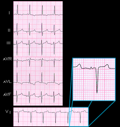
This ECG is an example of a sustained ectopic atrial tachycardia. It is from a 71 year old female. The rate is 115/min and the P waves are abnormal, indicating that they arise from an ectopic focus. They are upright in leads I and aVL but biphasic (negative-positive) in leads II and aVF and inverted in lead III. In lead aVR they are positive-negative. The frontal plane axis of this P wave is -10 degrees. Note that in lead V1, the negative component of the P wave precedes its positive component.