Home
Understanding the Electrocardiogram

Main navigation

  • Home
  • Understanding ECGs
    • Introduction
User account menu
  • Log in

Breadcrumb

  1. Home
  2. Understanding the Electrocardiogram In Health & Disease
  3. 1.0.0 Generation of the ECG
  4. 1.6.0 Repolarization

1.6.7

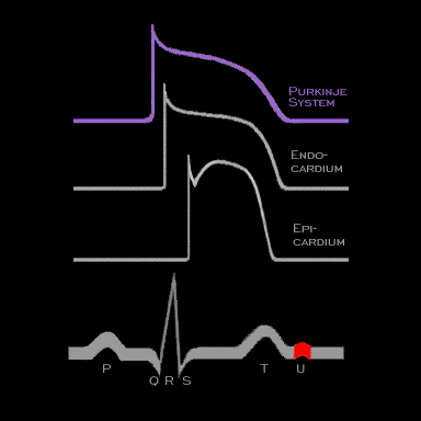
The T wave on the body surface is frequently followed by a small deflection called the U wave. The etiology of this wave remains unclear. Postulated mechanisms include the following: 1) repolarization of Purkinje fibers occurring after the ventricular fibers have repolarized; 2) delayed repolarization in some ventricular fibers; and 3) a wave generated in association with ventricular contraction, referred to as mechanico-electrical coupling. This latter mechanism is considered to be the most important of the three

Book traversal links for 1.6.7

  • 1.6.6
  • Up
  • 1.7.0 Events at the Cellular Level

Site is under construction

Book navigation

  • Introduction to First Edition
  • 1.0.0 Generation of the ECG
    • 1.1.0 Basic Physiology
    • 1.2.0 SA Node & Pacemakers
    • 1.3.0 Atrial Depolarization
    • 1.4.0 Atrio Ventricular Conduction
    • 1.5.0 Ventricular Depolarization
    • 1.6.0 Repolarization
      • 1.6.1
      • 1.6.2
      • 1.6.3
      • 1.6.4
      • 1.6.5
      • 1.6.6
      • 1.6.7
    • 1.7.0 Events at the Cellular Level
    • 1.8.0 Electrodes
    • 1.9.0 ECG Leads and Placement
  • 2.0.0 The Normal Electrocardiogram
  • 3.0.0 Inter and intra-ventricular Conduction Disturbances
  • 4.0.0 Ventricular Hypertrophy
  • 5.0.0 ELECTROLYTE ABNORMALITIES, DRUG EFFECTS AND THE LONG QT SYNDROMES
  • 6.0.0 Ischemia and Infarction - Introduction (frame i and ii)
  • 7.0.0 Tachycardias- Introduction
  • 8.0.0 The Bradycardias frame i-introduction
  • 9.0.0 The ECG of Heart Murmurs-introduction
  • 10.0.0 The Electrocardiogram in the Emergency Department-Introduction
Powered by Drupal