Home
Understanding the Electrocardiogram

Main navigation

  • Home
  • Understanding ECGs
    • Introduction
User account menu
  • Log in

Breadcrumb

  1. Home
  2. Understanding the Electrocardiogram In Health & Disease
  3. 1.0.0 Generation of the ECG
  4. 1.6.0 Repolarization

1.6.6

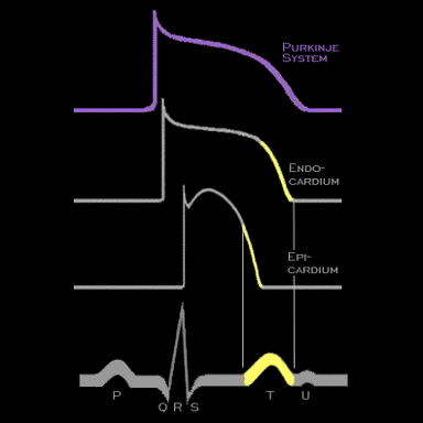
As one moves towards the epicardial surface, i.e. away from the endocardium, the action potentials first lengthen due to a unique population of cells in the mid-myocardium referred to as "M cells" and then shorten. The characteristics of rapid repolarization are reflected in the shape, duration and polarity of the T wave on the body surface electrocardiogram. As mentionhed earlier, the onset of the T wave coincides with the onset of rapid repolarization in those cells which are the first to repolarize while the end of the T wave coincides with the end of repolarization in those cells which are the last to repolarize.

Book traversal links for 1.6.6

  • 1.6.5
  • Up
  • 1.6.7

Site is under construction

Book navigation

  • Introduction to First Edition
  • 1.0.0 Generation of the ECG
    • 1.1.0 Basic Physiology
    • 1.2.0 SA Node & Pacemakers
    • 1.3.0 Atrial Depolarization
    • 1.4.0 Atrio Ventricular Conduction
    • 1.5.0 Ventricular Depolarization
    • 1.6.0 Repolarization
      • 1.6.1
      • 1.6.2
      • 1.6.3
      • 1.6.4
      • 1.6.5
      • 1.6.6
      • 1.6.7
    • 1.7.0 Events at the Cellular Level
    • 1.8.0 Electrodes
    • 1.9.0 ECG Leads and Placement
  • 2.0.0 The Normal Electrocardiogram
  • 3.0.0 Inter and intra-ventricular Conduction Disturbances
  • 4.0.0 Ventricular Hypertrophy
  • 5.0.0 ELECTROLYTE ABNORMALITIES, DRUG EFFECTS AND THE LONG QT SYNDROMES
  • 6.0.0 Ischemia and Infarction - Introduction (frame i and ii)
  • 7.0.0 Tachycardias- Introduction
  • 8.0.0 The Bradycardias frame i-introduction
  • 9.0.0 The ECG of Heart Murmurs-introduction
  • 10.0.0 The Electrocardiogram in the Emergency Department-Introduction
Powered by Drupal