Home
Understanding the Electrocardiogram

Main navigation

  • Home
  • Understanding ECGs
    • Introduction
User account menu
  • Log in

Breadcrumb

  1. Home
  2. Understanding the Electrocardiogram In Health & Disease
  3. 3.0.0 Inter and intra-ventricular Conduction Disturbances
  4. 3.2.0 Left Bundle Branch Block (LBBB)

3.2.5 (24)

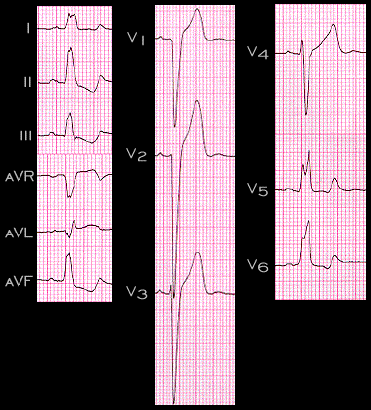
In some patients, usually those who are younger and without obvious underlying heart disease, the secondary changes in repolarization may be less pronounced. This ECG was recorded on an asymptomatic 50 year old physician with no underlying cardiovascular disease. Note that although there is ST segment depression and the T waves are abnormal, the abnormalities are less marked than on the prevous tracings.

Book traversal links for 3.2.5 (24)

  • 3.2.4 (23)
  • Up
  • 3.2.6 (25)

Site is under construction

Book navigation

  • Introduction to First Edition
  • 1.0.0 Generation of the ECG
  • 2.0.0 The Normal Electrocardiogram
  • 3.0.0 Inter and intra-ventricular Conduction Disturbances
    • 3.0.1 Introduction Continued
    • 3.1.0 Right Bundle Branch Block (RBBB)
    • 3.2.0 Left Bundle Branch Block (LBBB)
      • 3.2.1 (20)
      • 3.2.2 (21)
      • 3.2.3 (22)
      • 3.2.4 (23)
      • 3.2.5 (24)
      • 3.2.6 (25)
      • 3.2.7 (26)
      • 3.2.8 (28-frame 27 is omitted)
    • 3.3.0 Fascicular blocks (frame 29)
    • 3.4.0 Aberrant Ventricular Conduction (52)
    • 3.5.0 Ventricular Pre-excitation (77)
  • 4.0.0 Ventricular Hypertrophy
  • 5.0.0 ELECTROLYTE ABNORMALITIES, DRUG EFFECTS AND THE LONG QT SYNDROMES
  • 6.0.0 Ischemia and Infarction - Introduction (frame i and ii)
  • 7.0.0 Tachycardias- Introduction
  • 8.0.0 The Bradycardias frame i-introduction
  • 9.0.0 The ECG of Heart Murmurs-introduction
  • 10.0.0 The Electrocardiogram in the Emergency Department-Introduction
Powered by Drupal