
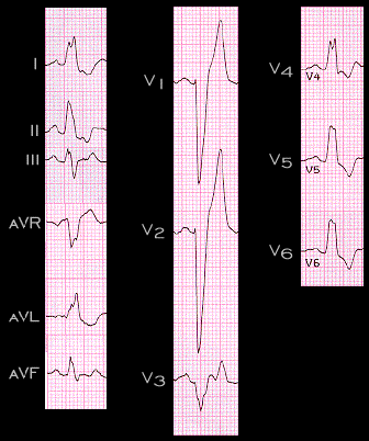
The ECG shown here, recorded from a 75 year old woman, is an example of typical complete left bundle branch block. Note that there are no small initial (septal) Q waves in leads 1, aVL or V6 (the left sided leads), that the QRS complex is diffusely widened and that there is a prominent notch in the QRS complex in leads I, aVL V4 and V5. Note also that the time from the onset of the QRS complex to the second peak of the R wave in the leads with the prominent notch is delayed, exceeding 60 ms and that the T wave is inverted in the leads where it is normally upright, i.e. leads I, II, V4, V5, and V6. These T wave changes reflect the changes in the sequence of repolarization that are secondary to the changes in the sequence of depolarization.