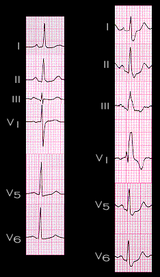
The persistence of the initial and middle portions of the QRS complex after the development of right bundle branch block is demonstrated here. The 2 ECGs shown here are from the same patient. The tracing on the left is normal. The tracing on the right was recorded one month later and shows complete right bundle branch block (RBBB). Note that the initial and mid portions of the QRS complex, i.e. those associated with septal and left ventricular depolarization, are the same in both tracings. Drawing the frontal plane QRS spatial vectors before and after the development of RBBB helps to illustrate this point. Try to do this before going to the next page.
