Home
Understanding the Electrocardiogram

Main navigation

  • Home
  • Understanding ECGs
    • Introduction
User account menu
  • Log in

Breadcrumb

  1. Home
  2. Understanding the Electrocardiogram In Health & Disease
  3. 3.0.0 Inter and intra-ventricular Conduction Disturbances
  4. 3.1.0 Right Bundle Branch Block (RBBB)

3.1.3

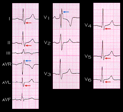
The ECG shown here illustrates the typical electrocardiographic features of complete right bundle branch block. It is important to note that  because impulse transmission through the left bundle is unaffected,  the  initial part of the QRS complex, i.e. a small Q wave in leads II and III and a small R wave in leads aVR and V1, which reflect the normal left-to-right depolarization of the interventricular septum is unchanged. The main portion of the QRS complex is also unchanged with R waves in leads I, II, aVF and V3-6 and S waves in leads aVR and V1. These reflect the normal sequence of left ventricular depolarization.

The unopposed right ventricular forces are represented by the broad S wave  in leads I and V6, i.e. the left sided and posterior/inferior leads (the red arrows), and by the R’ in leads aVR and V1, i.e. the right sided, and anterior/superior leads  (the blue arrows) . The duration of the QRS complex is prolonged and measures 0.12 sec.

Book traversal links for 3.1.3

  • 3.1.2
  • Up
  • 3.1.4

Site is under construction

Book navigation

  • Introduction to First Edition
  • 1.0.0 Generation of the ECG
  • 2.0.0 The Normal Electrocardiogram
  • 3.0.0 Inter and intra-ventricular Conduction Disturbances
    • 3.0.1 Introduction Continued
    • 3.1.0 Right Bundle Branch Block (RBBB)
      • 3.1.1
      • 3.1.2
      • 3.1.3
      • 3.1.4
      • 3.1.5
      • 3.1.6
      • 3.1.7
      • 3.1.8
      • 3.1.9
      • 3.1.10
      • 3.1.12. (14)
      • 3.1.13
      • 3.1.14
      • 3.1.15
      • 3.1.16
    • 3.2.0 Left Bundle Branch Block (LBBB)
    • 3.3.0 Fascicular blocks (frame 29)
    • 3.4.0 Aberrant Ventricular Conduction (52)
    • 3.5.0 Ventricular Pre-excitation (77)
  • 4.0.0 Ventricular Hypertrophy
  • 5.0.0 ELECTROLYTE ABNORMALITIES, DRUG EFFECTS AND THE LONG QT SYNDROMES
  • 6.0.0 Ischemia and Infarction - Introduction (frame i and ii)
  • 7.0.0 Tachycardias- Introduction
  • 8.0.0 The Bradycardias frame i-introduction
  • 9.0.0 The ECG of Heart Murmurs-introduction
  • 10.0.0 The Electrocardiogram in the Emergency Department-Introduction
Powered by Drupal