Home
Understanding the Electrocardiogram

Main navigation

  • Home
  • Understanding ECGs
    • Introduction
User account menu
  • Log in

Breadcrumb

  1. Home
  2. Understanding the Electrocardiogram In Health & Disease
  3. 9.0.0 The ECG of Heart Murmurs-introduction
  4. 9.1.0 Systolic Heart Murmurs (frame 3)
  5. 9.1.10 (12)

9.1.18 (skip to frame 26)

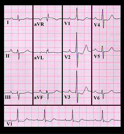
The ECG shown here is from a 54 year old slightly cyanotic woman with a soft, grade 2/6 systolic murmur at the left sternal border. Her ECG shows left axis deviation,  but the tall R wave and diminutive S wave in V1 and the S wave in V6 which is of greater amplitude than the R wave are indications of the right ventricular hypertrophy. This finding, in association with the soft murmur and the cyanosis suggests a ventricular septal defect with pulmonary hypertension and a balanced or bi-directional shunt (i.e. right to left as well as left to right). This is referred to as Eisenminger’s physiology. At cardiac catheterization, she was found to have a large ventricular septal defect with pulmonary hypertension, equalization of her right and left ventricular systolic pressures and a bi-directional shunt.

Book traversal links for 9.1.18 (skip to frame 26)

  • 9.1.17 (21-omit frames 18-20)
  • Up
  • 9.1.19 (frame 28) (frame 27 omitted)

Site is under construction

Book navigation

  • Introduction to First Edition
  • 1.0.0 Generation of the ECG
  • 2.0.0 The Normal Electrocardiogram
  • 3.0.0 Inter and intra-ventricular Conduction Disturbances
  • 4.0.0 Ventricular Hypertrophy
  • 5.0.0 ELECTROLYTE ABNORMALITIES, DRUG EFFECTS AND THE LONG QT SYNDROMES
  • 6.0.0 Ischemia and Infarction - Introduction (frame i and ii)
  • 7.0.0 Tachycardias- Introduction
  • 8.0.0 The Bradycardias frame i-introduction
  • 9.0.0 The ECG of Heart Murmurs-introduction
    • 9.0.1 (frame 1)
    • 9.0.2 (2)
    • 9.1.0 Systolic Heart Murmurs (frame 3)
      • 9.1.1 (4)
      • 9.1.2 (frame 4 again)
      • 9.1.3 (5)
      • 9.1.4 (6)
      • 9.1.5 (7)
      • 9.1.6 (8)
      • 9.1.7 (9)
      • 9.1.8 (10)
      • 9.1.9 (11)
      • 9.1.10 (12)
        • 9.1.11 (13)
        • 9.1.12 (14)
        • 9.1.13 (15)
        • 9.1.14 (16)
        • 9.1.15 (17)
        • 9.1.16 (18)
        • 9.1.17 (21-omit frames 18-20)
        • 9.1.18 (skip to frame 26)
        • 9.1.19 (frame 28) (frame 27 omitted)
      • 9.1.20 (29)
    • 9.2.0 Diastolic Murmurs (frame 38)
    • 9.3.0 Systolic and Diastolic Murmurs (frame 43)
  • 10.0.0 The Electrocardiogram in the Emergency Department-Introduction
Powered by Drupal