
As discussed earlier in this chapter, atrial fibrillation is caused by multiple small reentry circuits, or wavelets, that are usually located in the left atrium. The ventricular rhythm is characteristically irregular because of the variable penetration of the rapidly occurring fibrillatory waves into the AV node. These features are reflected on the body surface electrocardiogram, The atrial waves are usually less well defined than those associated with atrial flutter and the interval between the waves is shorter than that which occurs in atrial flutter because the rhythm is more rapid.
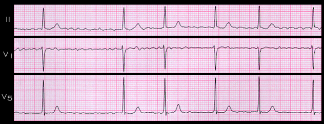
The ECG shown here, from a 35 year old male with acute mitral regurgitation secondary to a ruptured chordae tendineae, is an example of atrial fibrillation. At the time of this tracing, he was being treated with verapamil, a calcium channel blocking agent, to slow the ventricular rate. Note that there are no clearly defined P waves or flutter waves. Rather, there are fibrillatory waves of varying amplitude and rate. The QRS complexes are irregular with RR intervals that range from 0.82 to 1.56 seconds.