
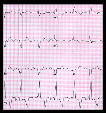
The ECG shown here also illustrates the difficulty in distinguishing slow atrial flutter from an ectopic atrial rhythm. The tracing is from a 45 year old male with severe congestive heart failure who was being treated with digoxin and hydrochlorthiazide. His serum potassium was 2.8 mm/L. The atrial rate is 200. There is 3:1 AV block and the ventricular rate is 68. The P waves are inverted in leads II, III and aVF and resemble type I atrial flutter waves with counterclockwise rotation. Although a diagnosis of slow atrial flutter with 3:1 AV block is not unreasonable, the more likely diagnosis is an ectopic atrial tachycardia with AV block, perhaps a manifestation of digitalis excess and hypopotassemia. The QRS complexes show right bundle branch block with left anterior fascicular block.