Home
Understanding the Electrocardiogram

Main navigation

  • Home
  • Understanding ECGs
    • Introduction
User account menu
  • Log in

Breadcrumb

  1. Home
  2. Understanding the Electrocardiogram In Health & Disease
  3. 7.0.0 Tachycardias- Introduction
  4. 7.3.0 Atrial Flutter and Fibrillation (frame 93)

7.3.2 (95)

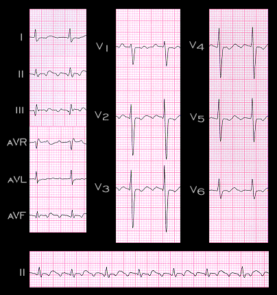
Another example of type I flutter is shown here. Again, the flutter waves are in inverted in leads II, III and aVF and upright in aVR, reflecting the superior direction of the atrial depolarization associated with the counterclockwise rotation of the impulse around the re-entry circuit. As in the previous example, the flutter rate is 300, but this time the ventricular rate is 100 because there is 3:1 AV block. The third flutter wave associated with each QRS complex is difficult to appreciate because it is concealed within the initial portion of the QRS complex.

Book traversal links for 7.3.2 (95)

  • 7.3.1 (94)
  • Up
  • 7.3.3 (96)

Site is under construction

Book navigation

  • Introduction to First Edition
  • 1.0.0 Generation of the ECG
  • 2.0.0 The Normal Electrocardiogram
  • 3.0.0 Inter and intra-ventricular Conduction Disturbances
  • 4.0.0 Ventricular Hypertrophy
  • 5.0.0 ELECTROLYTE ABNORMALITIES, DRUG EFFECTS AND THE LONG QT SYNDROMES
  • 6.0.0 Ischemia and Infarction - Introduction (frame i and ii)
  • 7.0.0 Tachycardias- Introduction
    • 7.1.0 Mechanisms of arrhythmias-frame 1
    • 7.2.0 Narrow Complex Rhythms (frame 38)
    • 7.3.0 Atrial Flutter and Fibrillation (frame 93)
      • 7.3.1 (94)
      • 7.3.2 (95)
      • 7.3.3 (96)
      • 7.3.4 (97)
      • 7.3.5 (98)
      • 7.3.6 (99)
      • 7.3.7 (100)
      • 7.3.8 (101)
      • 7.3.9 (102)
      • 7.3.10 (frame 103)
      • 7.3.20 (frame 113)
    • 7.4.0 Wide Complex Beats and Rhythms (frame 122)
  • 8.0.0 The Bradycardias frame i-introduction
  • 9.0.0 The ECG of Heart Murmurs-introduction
  • 10.0.0 The Electrocardiogram in the Emergency Department-Introduction
Powered by Drupal