Home
Understanding the Electrocardiogram

Main navigation

  • Home
  • Understanding ECGs
    • Introduction
User account menu
  • Log in

Breadcrumb

  1. Home
  2. Understanding the Electrocardiogram In Health & Disease
  3. 7.0.0 Tachycardias- Introduction
  4. 7.3.0 Atrial Flutter and Fibrillation (frame 93)

7.3.1 (94)

In atrial flutter, the atrial deflections seen on the body surface electrocardiogram are referred to as "f waves" or "flutter waves". They are characteristically rapid and regular with a saw-tooth configuration. In type I atrial flutter, the flutter rate is usually in the 250-350 per minute range and there is usually 2:1 AV block so that the ventricular rate is usually around 150 per minute. However, in younger individuals the AV nodal refractory period may be shorter, and capable of allowing 1:1 conduction, whereas in older individuals and those on a variety of cardioactive drugs, the AV nodal refractory period may be prolonged causing higher grades of AV block and a slower ventricular rate. The rhythm may be regular or irregular, depending on the consistency of the AV nodal block.

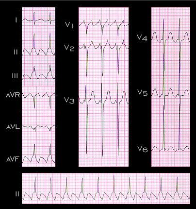
The tracing shown here is from a 71 year old male. It illustrates the most common ECG presentation of type I atrial flutter. The negative flutter waves in leads II, III and aVF reflect the fact that the counter-clockwise atrial depolarization in the anterior portion of the right atrial reentry circuit occurs in a caudal to cephalic (i.e.superior) direction. The rate of the flutter waves in this example is 300 per minute and there is 2:1 AV block which is constant. This results in a regular ventricular rate of 150 per minute. The increased R and S wave voltages in the precordial (chest) leads suggest LVH.

Book traversal links for 7.3.1 (94)

  • 7.3.0 Atrial Flutter and Fibrillation (frame 93)
  • Up
  • 7.3.2 (95)

Site is under construction

Book navigation

  • Introduction to First Edition
  • 1.0.0 Generation of the ECG
  • 2.0.0 The Normal Electrocardiogram
  • 3.0.0 Inter and intra-ventricular Conduction Disturbances
  • 4.0.0 Ventricular Hypertrophy
  • 5.0.0 ELECTROLYTE ABNORMALITIES, DRUG EFFECTS AND THE LONG QT SYNDROMES
  • 6.0.0 Ischemia and Infarction - Introduction (frame i and ii)
  • 7.0.0 Tachycardias- Introduction
    • 7.1.0 Mechanisms of arrhythmias-frame 1
    • 7.2.0 Narrow Complex Rhythms (frame 38)
    • 7.3.0 Atrial Flutter and Fibrillation (frame 93)
      • 7.3.1 (94)
      • 7.3.2 (95)
      • 7.3.3 (96)
      • 7.3.4 (97)
      • 7.3.5 (98)
      • 7.3.6 (99)
      • 7.3.7 (100)
      • 7.3.8 (101)
      • 7.3.9 (102)
      • 7.3.10 (frame 103)
      • 7.3.20 (frame 113)
    • 7.4.0 Wide Complex Beats and Rhythms (frame 122)
  • 8.0.0 The Bradycardias frame i-introduction
  • 9.0.0 The ECG of Heart Murmurs-introduction
  • 10.0.0 The Electrocardiogram in the Emergency Department-Introduction
Powered by Drupal