
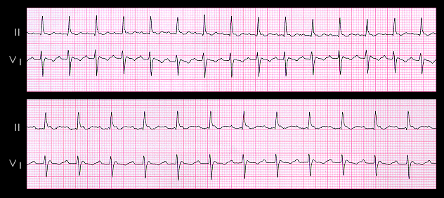
Earlier in this section, the difficulty in identifying flutter waves when they were embedded in the QRS complex was discussed. This becomes more of a problem when the flutter rate slows. The ECGs shown here are from an 85 year old female who was known to have persistent atrial flutter for many years. The two tracings were recorded 6 years apart and both show atrial flutter with 2:1 AV block. In the upper tracing the flutter rate is 270 and the ventricular rate is 135. In the lower, more recent tracing, the flutter rate has slowed to 220 and the ventricular rate is 110. In both tracings, one flutter wave is quite obvious but the other is located within the end of the QRS complex and is less obvious. The lower tracing, might have been misinterpreted as an ectopic atrial tachycardia with 1:1 AV conduction if the reader had failed to recognize the flutter wave superimposed on a the end of the QRS complex, or, if both flutter waves were recognized, as an even more rapid ectopic atrial tachycardia with 2:1 AV block.