Home
Understanding the Electrocardiogram

Main navigation

  • Home
  • Understanding ECGs
    • Introduction
User account menu
  • Log in

Breadcrumb

  1. Home
  2. Understanding the Electrocardiogram In Health & Disease
  3. 6.0.0 Ischemia and Infarction - Introduction (frame i and ii)
  4. 6.4.0 (Bundle Branch Blocks and othe IVCDs--frame 87)

6.4.12 (100)

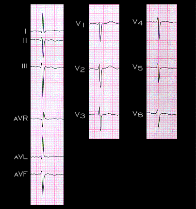
The electrocardiographic features of left anterior fascicular block may both mask and simulate the ECG findings of a prior Q wave infarction. The ECG shown here demonstrates the classical changes of left anterior fascicular block. As listed on page 3.3.2, these include:

  • Small Q wave in leads I, aVL, and/or V5 and V6
  • Small R wave in leads 3 and aVF.
  • Frontal plane QRS axis between -30 and -90 degrees, although some insist that it be between -45 and -90 degrees (it is -60 degrees in his racing).
  •  QRS duration of less than 0.12 seconds.
  • Poor R wave progression in chest leads with  S wave in leads V5 and V6

Book traversal links for 6.4.12 (100)

  • 6.4.11 (99)
  • Up
  • 6.4.13 (101)

Site is under construction

Book navigation

  • Introduction to First Edition
  • 1.0.0 Generation of the ECG
  • 2.0.0 The Normal Electrocardiogram
  • 3.0.0 Inter and intra-ventricular Conduction Disturbances
  • 4.0.0 Ventricular Hypertrophy
  • 5.0.0 ELECTROLYTE ABNORMALITIES, DRUG EFFECTS AND THE LONG QT SYNDROMES
  • 6.0.0 Ischemia and Infarction - Introduction (frame i and ii)
    • 6.1.0 ST Segment (frame1)
    • 6.2.0 Q WAVES (frame 53)
    • 6.3.0 T WAVES (frame 75)
    • 6.4.0 (Bundle Branch Blocks and othe IVCDs--frame 87)
      • 6.4.1 (88)
      • 6.4.2 (89)
      • 6.4.3 (frame 90)
      • 6.4.4 (91)
      • 6.4.5 (92)
      • 6.4.6 (93)
      • 6.4.7 (95) (omit 94)
      • 6.4.8 (96)
      • 6.4.9 (97)
      • 6.4.10 (frame 98)
      • 6.4.11 (99)
      • 6.4.12 (100)
      • 6.4.13 (101)
      • 6.4.14 (102)
      • 6.4.15 (103)
      • 6.4.16 (104)
      • 6.4.17 (105)
    • 6.5.0 (frame 106) FALSE POSITIVES
  • 7.0.0 Tachycardias- Introduction
  • 8.0.0 The Bradycardias frame i-introduction
  • 9.0.0 The ECG of Heart Murmurs-introduction
  • 10.0.0 The Electrocardiogram in the Emergency Department-Introduction
Powered by Drupal