
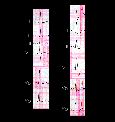
The secondary changes in repolarization are opposite to the changes in depolarization and are directed posteriorly and to the left. This is the same direction as the normal T wave vector and results in a slight exaggeration of the normal T wave. This is shown here. The ECGs are the same as those shown on the previous page. The T wave in the setting of RBBB (red arrow) is more prominent in leads I, V5 and V6, and is now inverted in lead V1. Thus, in the setting of RBBB, the changes in ST segment and the T wave changes associated with ischemia are essentially the same as those occurring in the absence of RBBB.