
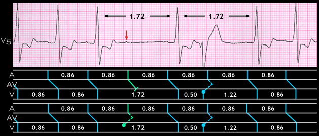
The 1.72 second pause containing the non-conducted P wave has 2 possible explanations. The first is that it may be due to type II 2nd degree AV block. This possibility is depicted in the ladder diagram above the ECG strip.
The alternative possible explanation is that it is caused by a concealed early ventricular premature beat that does not activate the ventricular myocardium because of “exit bock”. However, it is able to penetrate the AV junction in a retrograde direction to render it refractory when the sinus beat arrives. This possibility is depicted in the ladder diagram below the ECG strip and is supported by the presence of a manifest ventricular beat that obscures the sinus P wave. Note that the interval between the QRS complexes that precede and follow the ventricular premature beat, and the RR interval that encompasses the non-conducted P wave are both 1.76 seconds in duration, twice the sinus interval of 0.86 seconds.