Home
Understanding the Electrocardiogram

Main navigation

  • Home
  • Understanding ECGs
    • Introduction
User account menu
  • Log in

Breadcrumb

  1. Home
  2. Understanding the Electrocardiogram In Health & Disease
  3. 8.0.0 The Bradycardias frame i-introduction
  4. 8.3.0 Group Beating (frame 57)

8.3.5


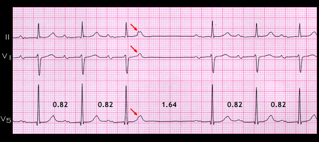
As mentioned, the interval of 1.64 seconds between the QRS complexes that precede and follows the non-conducted atrial premature beat (solid arrow) is exactly twice the sinus RR (or PP) interval, i.e., a fully compensatory pause. This is unusual for an atrial premature beats (see pages 7.4.4 and 7.4.5) because it will usually travel to the sinus node and reset it with the result that the post extrasystolic pause will most often be less than twice the normal RR interval, (i.e. non-compensatory). The most likely postulate to explain the pause in this patient is that because of the prolonged PR interval, the sinus discharge- shown here by the dotted arrow- occurred before the non-conducted atrial premature beat traveled to the sinus node to reset it. However, this sinus node impulse did not depolarize the atria to cause a P wave because the atria, having just been depolarized by the atrial premature beat, were still refractory. Hence, the fully compensatory pause.

Book traversal links for 8.3.5

  • 8.3.4 (61)
  • Up
  • 8.3.6 (62)

Site is under construction

Book navigation

  • Introduction to First Edition
  • 1.0.0 Generation of the ECG
  • 2.0.0 The Normal Electrocardiogram
  • 3.0.0 Inter and intra-ventricular Conduction Disturbances
  • 4.0.0 Ventricular Hypertrophy
  • 5.0.0 ELECTROLYTE ABNORMALITIES, DRUG EFFECTS AND THE LONG QT SYNDROMES
  • 6.0.0 Ischemia and Infarction - Introduction (frame i and ii)
  • 7.0.0 Tachycardias- Introduction
  • 8.0.0 The Bradycardias frame i-introduction
    • 8.1.0 Sinus Bradycardia and AV block (frame 1)
    • 8.2.0 SA Block (frame 41)
    • 8.3.0 Group Beating (frame 57)
      • 8.3.1 (58)
      • 8.3.2 (59)
      • 8.3.3 (60)
      • 8.3.4 (61)
      • 8.3.5
      • 8.3.6 (62)
      • 8.3.7 (63)
      • 8.3.8 (64)
      • 8.3.9 (65)
      • 8.3.10 (66)
      • 8.3.11 (67)
      • 8.3.12
  • 9.0.0 The ECG of Heart Murmurs-introduction
  • 10.0.0 The Electrocardiogram in the Emergency Department-Introduction
Powered by Drupal