Home
Understanding the Electrocardiogram

Main navigation

  • Home
  • Understanding ECGs
    • Introduction
User account menu
  • Log in

Breadcrumb

  1. Home
  2. Understanding the Electrocardiogram In Health & Disease
  3. 7.0.0 Tachycardias- Introduction
  4. 7.4.0 Wide Complex Beats and Rhythms (frame 122)
  5. 7.4.40 (frame 163)

7.4.41 (164)

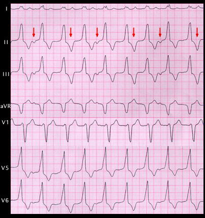
[THE 2nd 4th AND 6th ARROWS SHOULD BE BROKEN (RATHER THAN SOLID)]

The ECG is characteristic of the ventricular tachycardias that originate in the right ventricular outflow tract and are labeled as such, i.e. right ventricular outflow tract (RVOT) tachycardia. These arrhythmias are catecholamine sensitive, are often brought on by exercise and frequently respond to treatment with beat-adrenergic blocking agents. The retrograde P waves are marked. The solid arrows point at obvious P waves. The broken arrows point at T waves that are a bit different than their neighbors, suggesting that they contain an additional P wave having a VA interval that is less than that associated with the following, obvious P wave. This complicated relationship of the superiorly directed P waves to the QRS complexes suggests 3:2 retrograde VA conduction.

Book traversal links for 7.4.41 (164)

  • 7.4.40 (frame 163)
  • Up
  • 7.4.42 (165)

Site is under construction

Book navigation

  • Introduction to First Edition
  • 1.0.0 Generation of the ECG
  • 2.0.0 The Normal Electrocardiogram
  • 3.0.0 Inter and intra-ventricular Conduction Disturbances
  • 4.0.0 Ventricular Hypertrophy
  • 5.0.0 ELECTROLYTE ABNORMALITIES, DRUG EFFECTS AND THE LONG QT SYNDROMES
  • 6.0.0 Ischemia and Infarction - Introduction (frame i and ii)
  • 7.0.0 Tachycardias- Introduction
    • 7.1.0 Mechanisms of arrhythmias-frame 1
    • 7.2.0 Narrow Complex Rhythms (frame 38)
    • 7.3.0 Atrial Flutter and Fibrillation (frame 93)
    • 7.4.0 Wide Complex Beats and Rhythms (frame 122)
      • 7.4.1 (123)
      • 7.4.2 (124)
      • 7.4.3 (125)
      • 7.4.4 (126)
      • 7.4.5 (127)
      • 7.4.6 (128)
      • 7.4.7 (129)
      • 7.4.8 (130)
      • 7.4.9 (131)
      • 7.4.10 (frame 132)
      • 7.4.20 (frame 142)
      • 7.4.30 (frame 154)
      • 7.4.40 (frame 163)
        • 7.4.41 (164)
        • 7.4.42 (165)
        • 7.4.43 (166)
        • 7.4.44 (167)
        • 7.4.45 (new frame)
  • 8.0.0 The Bradycardias frame i-introduction
  • 9.0.0 The ECG of Heart Murmurs-introduction
  • 10.0.0 The Electrocardiogram in the Emergency Department-Introduction
Powered by Drupal