Home
Understanding the Electrocardiogram

Main navigation

  • Home
  • Understanding ECGs
    • Introduction
User account menu
  • Log in

Breadcrumb

  1. Home
  2. Understanding the Electrocardiogram In Health & Disease
  3. 1.0.0 Generation of the ECG
  4. 1.7.0 Events at the Cellular Level

1.7.1

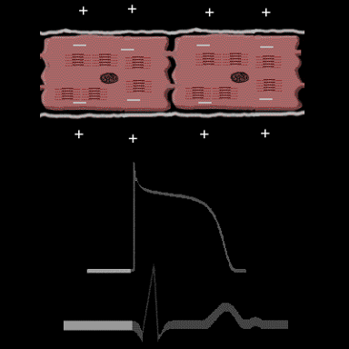
We know that when the heart is in its resting state, the inside of each individual cell is negative relative to the outside. This is referred to as the "resting membrane potential" (RMP) and is responsible for the resting phase (phase 4) of the action potential. In the normal heart there are no voltage gradients or electrical boundaries between the ventricular myocardial cells. No current flows between the cells, and there is no deflection on the body surface electrocardiogram, only a straight line.

Book traversal links for 1.7.1

  • 1.7.0 Events at the Cellular Level
  • Up
  • 1.7.2

Site is under construction

Book navigation

  • Introduction to First Edition
  • 1.0.0 Generation of the ECG
    • 1.1.0 Basic Physiology
    • 1.2.0 SA Node & Pacemakers
    • 1.3.0 Atrial Depolarization
    • 1.4.0 Atrio Ventricular Conduction
    • 1.5.0 Ventricular Depolarization
    • 1.6.0 Repolarization
    • 1.7.0 Events at the Cellular Level
      • 1.7.1
      • 1.7.2
      • 1.7.3
      • 1.7.4
      • 1.7.5
      • 1.7.6
      • 1.7.7
      • 1.7.8
      • 1.7.9
      • 1.7.10
      • 1.7.11
      • 1.7.12
    • 1.8.0 Electrodes
    • 1.9.0 ECG Leads and Placement
  • 2.0.0 The Normal Electrocardiogram
  • 3.0.0 Inter and intra-ventricular Conduction Disturbances
  • 4.0.0 Ventricular Hypertrophy
  • 5.0.0 ELECTROLYTE ABNORMALITIES, DRUG EFFECTS AND THE LONG QT SYNDROMES
  • 6.0.0 Ischemia and Infarction - Introduction (frame i and ii)
  • 7.0.0 Tachycardias- Introduction
  • 8.0.0 The Bradycardias frame i-introduction
  • 9.0.0 The ECG of Heart Murmurs-introduction
  • 10.0.0 The Electrocardiogram in the Emergency Department-Introduction
Powered by Drupal